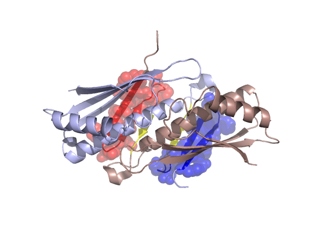
Structural Details of PDB entry 1VLA
Structural Details of PDB entry 1VLA
PDBid Chains Hinge Swapped Domain
1VLA
A,B
A:19-24,B:19-24
A:0-18,B:0-18
Swapped-domain interface residues and interactions:
Chains Residues
A
0 , 2 , 3 , 4 , 5 , 8 , 11 , 13 , 15 , 16 , 17 , 36 , 40 , 44 , 48 , 69 , 70 , 71 , 72 , 73 , 74 , 75 , 76 ,
B
0 , 2 , 3 , 4 , 5 , 8 , 11 , 13 , 15 , 16 , 17 , 36 , 40 , 48 , 69 , 70 , 71 , 72 , 73 , 74 , 75 , 76 ,
Non-swapped-domain interface residues and interactions:
Chains Residues
A
4 , 3 , 2 , 1 , 19 , 21 , 29 , 30 , 33 , 34 , 35 , 37 , 38 , 39 , 41 , 42 , 45 , 49 , 51 , 55 , 56 , 59 , 64 , 68 , 77 , 78 , 82 , 83 , 84 , 85 , 86 , 116 , 117 , 118 , 121 , 122 , 123 , 124 , 125 , 138 ,
B
3 , 1 , 19 , 21 , 29 , 30 , 33 , 34 , 35 , 37 , 38 , 39 , 41 , 42 , 44 , 45 , 49 , 51 , 55 , 56 , 59 , 64 , 68 , 77 , 78 , 80 , 82 , 83 , 84 , 85 , 86 , 116 , 117 , 118 , 121 , 122 , 123 , 124 , 125 ,