Home BrowseSearch Help Group

Pfam Domains mapped on to the structure: 1VL7

No. Chain ID Pfam ID Pfam Description Linkout - Pfam Linkout - CDD
1 A PF01243 Pyridoxamine 5'-phosphate oxidase PF01243 PF01243

Gene Ontology Annotations: 1VL7

No. GO ID GO Description Linkout - AmiGO
1 GO:0055114 oxidation-reduction process GO:0055114

Conserved Domain Database Superfamily Annotations: 1VL7

No. PDB ID PSSM ID CDD Accession Superfamily Short Name Linkout - CDD
1 1VL7 212207 cl00381 PNPOx_like superfamily - -
Structural Details of PDB entry 1VL7

Structural Details of PDB entry 1VL7

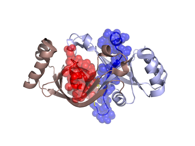
PDBid Chains Hinge Swapped Domain
1VL7 A,B A:31-32,B:31-32 A:11-30,B:11-30

Swapped-domain interface residues and interactions:

Chains Residues
A 18, 19, 20, 22, 24, 26, 30, 31, 32, 33, 34, 88,
B 18, 19, 20, 22, 24, 26, 30, 32, 33, 34,

Non-swapped-domain interface residues and interactions:

Chains Residues
A 35, 36, 38, 52, 54, 55, 66, 68, 70, 72, 74, 75, 76, 80, 81, 82, 84, 86, 90, 114, 117, 121, 127, 143, 145,
B 31, 35, 36, 38, 52, 54, 55, 66, 68, 70, 72, 74, 75, 76, 80, 81, 82, 84, 86, 88, 90, 114, 117, 121, 143,

Swapped domains are represented using trasperent spheres.
Non-swapped part is represented using light color and cartoon representation.
Hinge region is shown in yellow color.

Mutations in critical regions:

Chains Hinge Domain swapped interface Non-swapped interface Swapped Domain
ANo mutationNo mutationNo mutationNo mutation
BNo mutationNo mutationNo mutationNo mutation

HIDE output:

Homologues found through HIDE algorithm

JMOL Visualization:

Jmol_logo            Jmol_logo            Jmol_logo

2D-plot:

A:1VL7 B:1VL7

JOY Structural annotation for hinge hinge and swapped domain:

Hinge Swapped domain

JOY output:

ali file:1VL7.ali
atm file:1VL7.atm
cof file:1VL7.cof
hbd file:1VL7.hbd
html file:1VL7.html
pdb file:1VL7.pdb
ps file:1VL7.ps
psa file:1VL7.psa
rtf file:1VL7.rtf
sst file:1VL7.sst
tem file:1VL7.tem