Pfam Domains mapped on to the structure: 1VL2
No.
Chain ID
Pfam ID
Pfam Description
Linkout - Pfam
Linkout - CDD
1
A
PF00764
Arginosuccinate synthase
PF00764
PF00764
Gene Ontology Annotations: 1VL2
Conserved Domain Database Superfamily Annotations: 1VL2
Structural Details of PDB entry 1VL2
Structural Details of PDB entry 1VL2
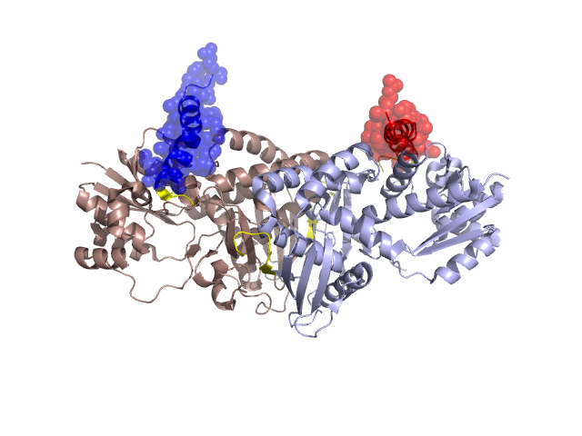
PDBid Chains Hinge Swapped Domain
1VL2
A,B
A:359-383,B:359-383
A:384-409,B:384-409
Swapped-domain interface residues and interactions:
Chains Residues
A
118 , 119 , 124 , 128 , 131 , 132 , 133 , 135 , 144 , 272 , 326 , 384 , 386 , 387 , 389 , 390 , 393 , 394 , 396 , 397 , 398 , 400 , 401 , 402 , 409 ,
B
118 , 119 , 124 , 128 , 131 , 132 , 133 , 135 , 144 , 326 , 384 , 386 , 387 , 389 , 390 , 393 , 394 , 396 , 397 , 398 , 400 , 401 , 402 , 404 ,
Non-swapped-domain interface residues and interactions:
Chains Residues
A
82 , 83 , 84 , 127 , 136 , 146 , 149 , 194 , 196 , 197 , 199 , 200 , 211 , 212 , 216 , 220 , 263 , 265 , 267 , 269 , 270 , 271 , 273 , 275 , 277 , 293 , 294 , 297 , 298 , 300 , 302 , 305 , 306 , 309 , 310 , 313 , 314 , 317 , 323 , 327 , 328 , 330 , 348 , 352 , 353 , 354 , 355 , 357 , 358 , 359 , 360 , 361 , 362 , 368 , 379 , 381 , 383 ,
B
82 , 83 , 84 , 127 , 136 , 146 , 149 , 194 , 196 , 197 , 199 , 200 , 211 , 212 , 216 , 220 , 263 , 265 , 267 , 269 , 270 , 271 , 272 , 273 , 275 , 293 , 294 , 297 , 298 , 300 , 302 , 305 , 306 , 309 , 310 , 313 , 314 , 317 , 323 , 327 , 328 , 330 , 348 , 352 , 353 , 354 , 355 , 357 , 358 , 359 , 360 , 361 , 362 , 368 , 381 , 382 , 383 ,
Mutations in critical regions:
Chains
Hinge
Domain swapped interface Non-swapped interface Swapped Domain
A No mutation No mutation No mutation No mutation B No mutation No mutation No mutation No mutation
HIDE output:
JMOL Visualization:
2D-plot:
JOY Structural annotation for hinge hinge and swapped domain:
JOY output: