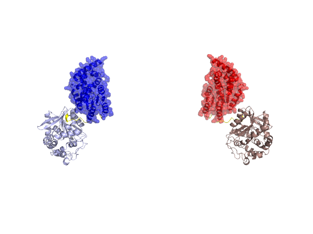
Pfam Domains mapped on to the structure: 1VJ5 No. Chain ID Pfam ID Pfam Description Linkout - Pfam Linkout - CDD 1 A PF13419 Haloacid dehalogenase-like hydrolase PF13419 PF13419 2 A PF12695 Alpha/beta hydrolase family PF12695 PF12695 3 A PF12697 Alpha/beta hydrolase family PF12697 PF12697 4 A PF00561 alpha/beta hydrolase fold PF00561 PF00561 Conserved Domain Database Superfamily Annotations: 1VJ5 No. PDB ID PSSM ID CDD Accession Superfamily Short Name Linkout - CDD 1 1VJ5 119389 HAD_like N cl11391 2 1VJ5 212620 HAD_like superfamily N - 3 1VJ5 211462 Esterase_lipase superfamily NC - 4 1VJ5 211462 Esterase_lipase superfamily NC - 5 1VJ5 211462 Esterase_lipase superfamily NC - 6 1VJ5 211462 Esterase_lipase superfamily NC - Structural Details of PDB entry 1VJ5 Structural Details of PDB entry 1VJ5 PDBid Chains Hinge Swapped Domain 1VJ5 A,B A:219-234,B:219-234 A:2-218,B:2-218 Swapped-domain interface residues and interactions: Chains Residues Non-swapped-domain interface residues and interactions: Chains Residues A 547, Swapped domains are represented using trasperent spheres. Non-swapped part is represented using light color and cartoon representation. Hinge region is shown in yellow color. Mutations in critical regions: Chains Hinge Domain swapped interface Non-swapped interface Swapped Domain ANo mutationNo mutationNo mutationNo mutation BNo mutationNo mutationNo mutationNo mutation HIDE output: Homologues found through HIDE algorithm JMOL Visualization: 2D-plot: A:1VJ5 B:1VJ5 JOY Structural annotation for hinge hinge and swapped domain: Hinge Swapped domain JOY output: ali file:1VJ5.ali atm file:1VJ5.atm cof file:1VJ5.cof hbd file:1VJ5.hbd html file:1VJ5.html pdb file:1VJ5.pdb ps file:1VJ5.ps psa file:1VJ5.psa rtf file:1VJ5.rtf sst file:1VJ5.sst tem file:1VJ5.tem