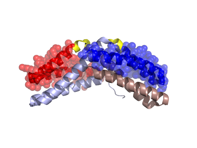
Pfam Domains mapped on to the structure: 1VH6
No.
Chain ID
Pfam ID
Pfam Description
Linkout - Pfam
Linkout - CDD
1
A
PF02561
Flagellar protein FliS
PF02561
PF02561
Gene Ontology Annotations: 1VH6
Conserved Domain Database Superfamily Annotations: 1VH6
Structural Details of PDB entry 1VH6
Structural Details of PDB entry 1VH6
PDBid Chains Hinge Swapped Domain
1VH6
A,B
A:75-79,B:75-79
A:80-119,B:80-122
Swapped-domain interface residues and interactions:
Chains Residues
A
20 , 21 , 25 , 28 , 32 , 35 , 39 , 42 , 51 , 54 , 58 , 61 , 65 , 82 , 83 , 86 , 89 , 90 , 93 , 96 , 98 , 101 , 104 , 105 , 108 , 109 , 111 , 112 , 115 , 116 , 119 ,
B
21 , 25 , 28 , 32 , 35 , 39 , 42 , 51 , 54 , 58 , 61 , 65 , 82 , 83 , 86 , 89 , 90 , 93 , 96 , 98 , 101 , 104 , 105 , 108 , 109 , 111 , 112 , 115 , 116 , 119 ,
Non-swapped-domain interface residues and interactions:
Chains Residues
A
18 , 19 , 23 , 24 , 27 , 29 , 31 , 36 , 43 , 50 , 52 , 55 , 56 , 59 , 62 , 63 , 64 , 66 , 67 , 68 , 71 , 72 , 74 , 75 , 76 , 79 ,
B
15 , 16 , 18 , 19 , 20 , 23 , 24 , 27 , 29 , 31 , 36 , 43 , 50 , 59 , 62 , 63 , 64 , 66 , 67 , 68 , 69 , 71 , 72 , 74 , 75 , 76 , 79 ,
Mutations in critical regions:
Chains
Hinge
Domain swapped interface Non-swapped interface Swapped Domain
A No mutation No mutation No mutation No mutation B No mutation No mutation No mutation No mutation
HIDE output:
JMOL Visualization:
2D-plot:
JOY Structural annotation for hinge hinge and swapped domain:
JOY output: