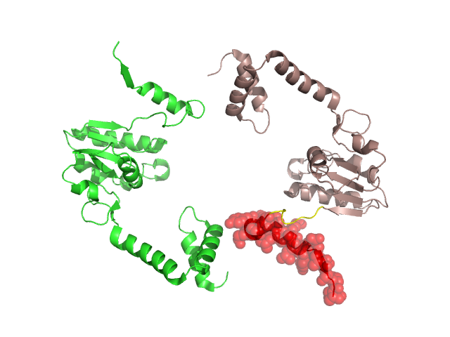
Pfam Domains mapped on to the structure: 1VDD No. Chain ID Pfam ID Pfam Description Linkout - Pfam Linkout - CDD 1 A PF02132 RecR protein PF02132 PF02132 2 A PF13662 Toprim domain PF13662 PF13662 3 A PF01751 Toprim domain PF01751 PF01751 Conserved Domain Database Superfamily Annotations: 1VDD No. PDB ID PSSM ID CDD Accession Superfamily Short Name Linkout - CDD 1 1VDD 173775 cd01025 TOPRIM_recR - cl00718 2 1VDD 207177 cl00718 TOPRIM superfamily - - 3 1VDD 202123 cl03461 RecR superfamily - - 4 1VDD 202123 cl03461 RecR superfamily - - Structural Details of PDB entry 1VDD Structural Details of PDB entry 1VDD PDBid Chains Hinge Swapped Domain 1VDD A,B A:168-175,B:168-175 A:176-199,B:176-300 Swapped-domain interface residues and interactions: Chains Residues A 179, 180, 183, 199, Non-swapped-domain interface residues and interactions: Chains Residues A 23, 26, 27, D 23, 26, 27, 179, 180, Swapped domains are represented using trasperent spheres. Non-swapped part is represented using light color and cartoon representation. Hinge region is shown in yellow color. Mutations in critical regions: Chains Hinge Domain swapped interface Non-swapped interface Swapped Domain ANo mutationNo mutationNo mutationNo mutation BNo mutationNo mutationNo mutationNo mutation HIDE output: Homologues found through HIDE algorithm JMOL Visualization: 2D-plot: A:1VDD D:1VDD JOY Structural annotation for hinge hinge and swapped domain: Hinge Swapped domain JOY output: ali file:1VDD.ali atm file:1VDD.atm cof file:1VDD.cof hbd file:1VDD.hbd html file:1VDD.html pdb file:1VDD.pdb ps file:1VDD.ps psa file:1VDD.psa rtf file:1VDD.rtf sst file:1VDD.sst tem file:1VDD.tem