Pfam Domains mapped on to the structure: 1V8D
No.
Chain ID
Pfam ID
Pfam Description
Linkout - Pfam
Linkout - CDD
1
A
PF04260
Protein of unknown function (DUF436)
PF04260
PF04260
Conserved Domain Database Superfamily Annotations: 1V8D
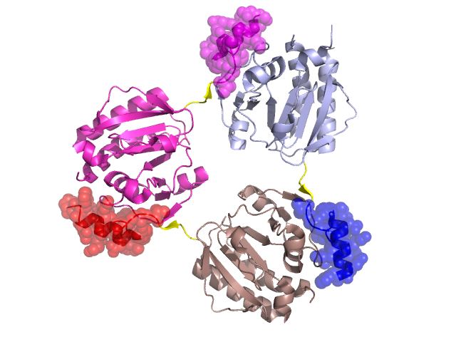
Structural Details of PDB entry 1V8D
Structural Details of PDB entry 1V8D
PDBid Chains Hinge Swapped Domain
1V8D
A,D,C,B
A:207-210,D:207-210,C:207-210,B:207-210
A:211-232,D:211-232,C:211-232,B:211-232
Swapped-domain interface residues and interactions:
Chains Residues
A
114 , 115 , 116 , 134 , 135 , 136 , 137 , 138 , 139 , 142 , 211 , 212 , 213 , 214 , 215 , 216 , 217 , 218 , 219 , 220 , 223 , 226 , 227 , 229 , 230 , 231 , 232 ,
B
114 , 115 , 116 , 134 , 135 , 136 , 137 , 138 , 139 , 142 , 211 , 212 , 213 , 214 , 215 , 216 , 217 , 218 , 219 , 220 , 223 , 226 , 227 , 229 ,
C
114 , 115 , 116 , 134 , 135 , 136 , 137 , 138 , 139 , 142 , 211 , 212 , 213 , 214 , 215 , 216 , 217 , 218 , 219 , 220 , 223 , 226 , 227 ,
Non-swapped-domain interface residues and interactions:
Chains Residues
A
83 , 84 , 86 , 132 , 133 , 140 , 141 , 148 , 151 , 152 , 157 , 184 , 208 , 209 , 210 ,
B
83 , 84 , 86 , 87 , 132 , 133 , 140 , 141 , 148 , 151 , 152 , 161 , 183 , 208 , 209 , 210 ,
C
86 , 132 , 133 , 140 , 141 , 148 , 151 , 152 , 161 , 184 , 208 , 209 , 210 ,
Mutations in critical regions:
Chains
Hinge
Domain swapped interface Non-swapped interface Swapped Domain
A No mutation No mutation No mutation No mutation D No mutation No mutation No mutation No mutation C No mutation No mutation No mutation No mutation B No mutation No mutation No mutation No mutation
HIDE output:
JMOL Visualization:
2D-plot:
JOY Structural annotation for hinge hinge and swapped domain:
JOY output: