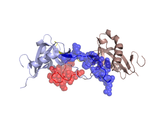
Structural Details of PDB entry 1V7P
Structural Details of PDB entry 1V7P
PDBid Chains Hinge Swapped Domain
1V7P
A,B
A:75-80,B:75-80;A:97-102,B:97-102
A:81-96,B:81-96
Swapped-domain interface residues and interactions:
Chains Residues
A
45 , 46 , 81 , 82 , 83 , 85 , 86 , 87 , 89 , 91 , 93 , 94 , 95 , 96 , 115 , 116 , 117 ,
B
40 , 42 , 43 , 66 , 67 , 70 , 71 , 72 , 73 , 74 , 81 , 82 , 84 , 86 , 88 , 89 , 90 , 91 , 106 , 107 , 108 ,
Non-swapped-domain interface residues and interactions:
Chains Residues
A
29 , 40 , 43 , 44 , 47 , 71 , 72 , 73 , 74 , 75 , 76 , 80 , 97 , 98 , 100 , 102 , 103 , 105 , 118 , 134 ,
B
37 , 41 , 44 , 47 , 75 , 76 , 77 , 78 , 79 , 80 , 104 , 105 , 109 ,