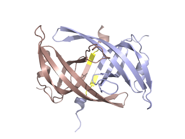
Structural Details of PDB entry 1V1Q
Structural Details of PDB entry 1V1Q
PDBid Chains Hinge Swapped Domain
1V1Q
A,B
A:002-4,B:002-4
A:4-1,B:4-1
Swapped-domain interface residues and interactions:
Non-swapped-domain interface residues and interactions:
Chains Residues
A
2 , 1 , 1 , 2 , 3 , 4 , 5 , 6 , 7 , 8 , 9 , 10 , 24 , 33 , 34 , 35 , 36 , 37 , 38 , 39 , 40 , 41 , 44 , 45 , 46 , 47 , 48 , 49 , 50 , 55 , 56 , 71 , 77 , 78 , 79 , 80 , 82 , 85 , 86 , 87 , 88 , 90 , 93 , 95 , 100 ,
B
7 , 4 , 2 , 1 , 1 , 2 , 3 , 4 , 5 , 6 , 7 , 8 , 9 , 10 , 33 , 34 , 35 , 36 , 37 , 38 , 39 , 40 , 42 , 44 , 46 , 47 , 48 , 49 , 51 , 55 , 56 , 58 , 71 , 77 , 78 , 79 , 80 , 81 , 82 , 87 , 88 , 89 , 90 , 93 , 95 , 100 ,