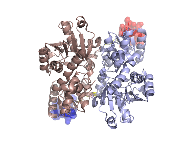
Pfam Domains mapped on to the structure: 1UIN
No.
Chain ID
Pfam ID
Pfam Description
Linkout - Pfam
Linkout - CDD
1
A
PF00291
Pyridoxal-phosphate dependent enzyme
PF00291
PF00291
Conserved Domain Database Superfamily Annotations: 1UIN
Structural Details of PDB entry 1UIN
Structural Details of PDB entry 1UIN
PDBid Chains Hinge Swapped Domain
1UIN
A,B
A:331-335,B:331-335
A:336-351,B:336-350
Swapped-domain interface residues and interactions:
Chains Residues
A
104 , 109 , 126 , 128 , 129 , 130 , 131 , 140 , 144 , 336 , 337 , 338 , 339 , 341 , 344 , 345 , 347 , 348 , 349 , 350 , 351 ,
B
104 , 109 , 126 , 128 , 129 , 130 , 131 , 140 , 144 , 336 , 337 , 338 , 339 , 341 , 344 , 345 , 347 , 348 , 349 ,
Non-swapped-domain interface residues and interactions:
Chains Residues
A
2 , 24 , 25 , 30 , 31 , 32 , 33 , 35 , 51 , 54 , 56 , 58 , 78 , 97 , 98 , 100 , 102 , 106 , 112 , 114 , 115 , 118 , 119 , 121 , 122 , 123 , 124 , 127 , 143 , 147 , 148 , 150 , 279 , 280 , 282 , 283 , 285 , 319 , 321 , 322 , 324 , 327 , 328 , 331 , 333 , 334 ,
B
24 , 25 , 28 , 30 , 32 , 33 , 35 , 51 , 54 , 56 , 58 , 97 , 98 , 100 , 102 , 106 , 114 , 115 , 118 , 119 , 121 , 122 , 123 , 124 , 127 , 139 , 143 , 147 , 148 , 150 , 174 , 279 , 280 , 282 , 283 , 285 , 319 , 321 , 322 , 324 , 327 , 328 , 330 , 331 , 332 , 333 , 334 ,
Mutations in critical regions:
Chains
Hinge
Domain swapped interface Non-swapped interface Swapped Domain
A No mutation No mutation No mutation No mutation B No mutation No mutation No mutation No mutation
HIDE output:
JMOL Visualization:
2D-plot:
JOY Structural annotation for hinge hinge and swapped domain:
JOY output: