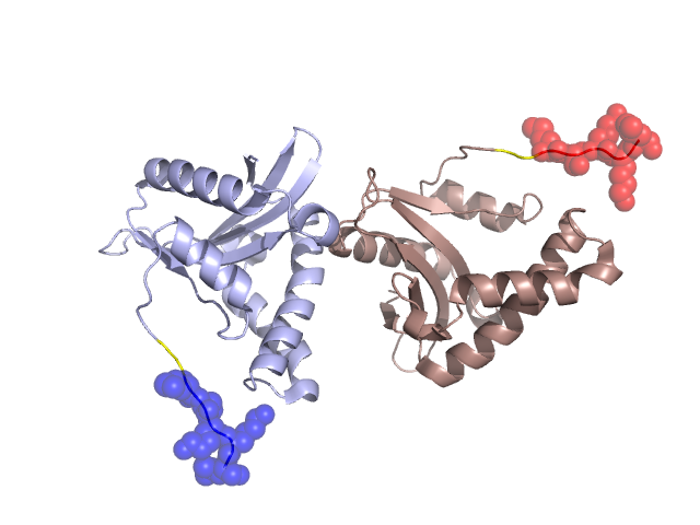
Pfam Domains mapped on to the structure: 1UFH No. Chain ID Pfam ID Pfam Description Linkout - Pfam Linkout - CDD 1 A PF13302 Acetyltransferase (GNAT) domain PF13302 PF13302 2 A PF13420 Acetyltransferase (GNAT) domain PF13420 PF13420 3 A PF13673 Acetyltransferase (GNAT) domain PF13673 PF13673 4 A PF13508 Acetyltransferase (GNAT) domain PF13508 PF13508 5 A PF00583 Acetyltransferase (GNAT) family PF00583 PF00583 6 A PF08445 FR47-like protein PF08445 PF08445 Conserved Domain Database Superfamily Annotations: 1UFH No. PDB ID PSSM ID CDD Accession Superfamily Short Name Linkout - CDD 1 1UFH 212199 cl00357 NAT_SF superfamily - - 2 1UFH 212199 cl00357 NAT_SF superfamily - - Structural Details of PDB entry 1UFH Structural Details of PDB entry 1UFH PDBid Chains Hinge Swapped Domain 1UFH A,B A:150-151,B:150-151 A:152-156,B:152-156 Swapped-domain interface residues and interactions: Chains Residues A 156, Non-swapped-domain interface residues and interactions: Chains Residues A 82, 83, 87, 115, 118, 119, 120, 121, 123, B 11, 14, 15, 18, 19, 70, 71, 97, 98, 99, 100, 103, Swapped domains are represented using trasperent spheres. Non-swapped part is represented using light color and cartoon representation. Hinge region is shown in yellow color. Mutations in critical regions: Chains Hinge Domain swapped interface Non-swapped interface Swapped Domain ANo mutationNo mutationNo mutationNo mutation BNo mutationNo mutationNo mutationNo mutation HIDE output: Homologues found through HIDE algorithm JMOL Visualization: 2D-plot: A:1UFH B:1UFH JOY Structural annotation for hinge hinge and swapped domain: Hinge Swapped domain JOY output: ali file:1UFH.ali atm file:1UFH.atm cof file:1UFH.cof hbd file:1UFH.hbd html file:1UFH.html pdb file:1UFH.pdb ps file:1UFH.ps psa file:1UFH.psa rtf file:1UFH.rtf sst file:1UFH.sst tem file:1UFH.tem