Home BrowseSearch Help Group

Pfam Domains mapped on to the structure: 1U6L

No. Chain ID Pfam ID Pfam Description Linkout - Pfam Linkout - CDD
1 A PF00903 Glyoxalase/Bleomycin resistance protein/Dioxygenase superfamily PF00903 PF00903

Conserved Domain Database Superfamily Annotations: 1U6L

No. PDB ID PSSM ID CDD Accession Superfamily Short Name Linkout - CDD
1 1U6L 176658 cd06588 PhnB_like - cl14632
2 1U6L 211465 cl14632 Glo_EDI_BRP_like superfamily - -
3 1U6L 211465 cl14632 Glo_EDI_BRP_like superfamily - -
Structural Details of PDB entry 1U6L

Structural Details of PDB entry 1U6L

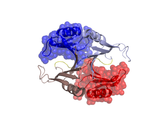
PDBid Chains Hinge Swapped Domain
1U6L A,B A:73-78,B:73-78 A:79-138,B:79-138

Swapped-domain interface residues and interactions:

Chains Residues
A 2, 3, 4, 5, 6, 7, 8, 9, 10, 77, 78, 79, 80, 81, 82, 83, 84, 85, 86, 87, 88, 89, 97, 115, 138,
B 2, 3, 4, 5, 6, 7, 8, 9, 10, 77, 78, 79, 80, 81, 82, 83, 84, 85, 86, 87, 88, 89, 115,

Non-swapped-domain interface residues and interactions:

Chains Residues
A 26, 40, 60, 62, 63, 65,
B 26, 40, 41, 60, 62, 63, 64, 65,

Swapped domains are represented using trasperent spheres.
Non-swapped part is represented using light color and cartoon representation.
Hinge region is shown in yellow color.

Mutations in critical regions:

Chains Hinge Domain swapped interface Non-swapped interface Swapped Domain
ANo mutationSER(2)-, LEU(3)-, No mutationNo mutation
BNo mutationSER(2)-, LEU(3)-, No mutationNo mutation

HIDE output:

Homologues found through HIDE algorithm

JMOL Visualization:

Jmol_logo            Jmol_logo            Jmol_logo

2D-plot:

A:1U6L B:1U6L

JOY Structural annotation for hinge hinge and swapped domain:

Hinge Swapped domain

JOY output:

ali file:1U6L.ali
atm file:1U6L.atm
cof file:1U6L.cof
hbd file:1U6L.hbd
html file:1U6L.html
pdb file:1U6L.pdb
ps file:1U6L.ps
psa file:1U6L.psa
rtf file:1U6L.rtf
sst file:1U6L.sst
tem file:1U6L.tem