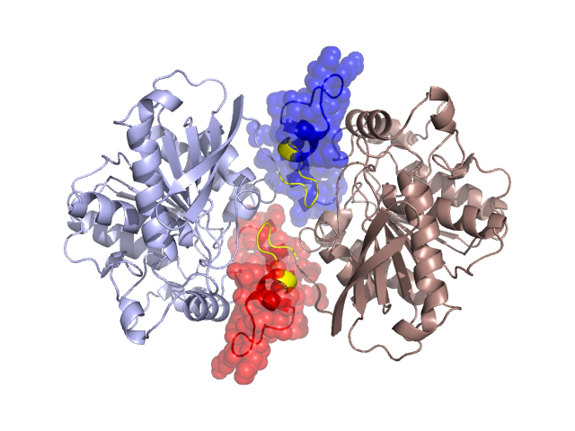
Structural Details of PDB entry 1U4N
Structural Details of PDB entry 1U4N
PDBid Chains Hinge Swapped Domain
1U4N
A,B
A:32-40,B:32-40
A:3-31,B:3-31
Swapped-domain interface residues and interactions:
Chains Residues
A
3 , 4 , 6 , 7 , 10 , 11 , 14 , 15 , 22 , 25 , 27 , 30 , 31 , 87 , 203 , 204 , 205 , 209 , 210 , 213 , 279 , 280 , 291 , 293 ,
B
3 , 4 , 6 , 7 , 10 , 11 , 14 , 15 , 22 , 25 , 27 , 30 , 31 , 87 , 203 , 204 , 205 , 209 , 210 , 213 , 279 , 280 , 291 , 293 ,
Non-swapped-domain interface residues and interactions:
Chains Residues
A
34 , 36 , 37 , 38 , 39 , 83 , 86 , 88 , 92 , 117 , 118 , 207 , 214 , 217 , 251 , 281 , 286 , 287 , 290 , 292 , 310 ,
B
34 , 36 , 37 , 38 , 39 , 83 , 86 , 88 , 92 , 117 , 118 , 207 , 214 , 217 , 251 , 281 , 286 , 287 , 290 , 292 ,