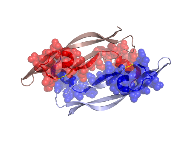
Structural Details of PDB entry 1TZH
Structural Details of PDB entry 1TZH
PDBid Chains Hinge Swapped Domain
1TZH
W,V
W:49-51,V:49-51
W:14-48,V:14-48
Swapped-domain interface residues and interactions:
Chains Residues
V
14 , 15 , 16 , 17 , 20 , 21 , 23 , 24 , 29 , 30 , 32 , 48 , 49 , 51 , 53 , 58 , 59 , 62 , 76 , 77 , 78 , 79 ,
W
14 , 15 , 16 , 17 , 20 , 21 , 23 , 24 , 29 , 30 , 32 , 48 , 49 , 51 , 53 , 58 , 59 , 62 , 76 , 77 , 78 , 79 ,
Non-swapped-domain interface residues and interactions:
Chains Residues
V
50 , 60 , 81 , 91 , 93 , 107 ,
W
50 , 60 , 81 , 91 , 93 ,