Home BrowseSearch Help Group

Pfam Domains mapped on to the structure: 1T92

No. Chain ID Pfam ID Pfam Description Linkout - Pfam Linkout - CDD
1 A PF08334 Type II secretion system (T2SS), protein G PF08334 PF08334

Conserved Domain Database Superfamily Annotations: 1T92

No. PDB ID PSSM ID CDD Accession Superfamily Short Name Linkout - CDD
1 1T92 192005 cl07082 T2SG superfamily - -
Structural Details of PDB entry 1T92

Structural Details of PDB entry 1T92

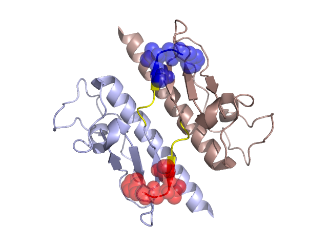
PDBid Chains Hinge Swapped Domain
1T92 A,B A:126-129,B:126-129 A:130-133,B:130-133

Swapped-domain interface residues and interactions:

Chains Residues
A 119, 120, 121, 122, 123, 130, 131, 132, 133,
B 119, 120, 121, 122, 123, 130, 131, 132,

Non-swapped-domain interface residues and interactions:

Chains Residues
A 30, 33, 34, 37, 38, 41, 44, 45, 48, 49, 52, 53, 101, 112, 124, 125, 128, 129,
B 30, 33, 34, 37, 38, 41, 44, 45, 48, 49, 52, 53, 101, 112, 124, 125, 128, 129,

Swapped domains are represented using trasperent spheres.
Non-swapped part is represented using light color and cartoon representation.
Hinge region is shown in yellow color.

Mutations in critical regions:

Chains Hinge Domain swapped interface Non-swapped interface Swapped Domain
ANo mutationPHE(133)-, No mutationPHE(133)-,
BNo mutationNo mutationNo mutationPHE(133)-,

HIDE output:

Homologues found through HIDE algorithm

JMOL Visualization:

Jmol_logo            Jmol_logo            Jmol_logo

2D-plot:

A:1T92 B:1T92

JOY Structural annotation for hinge hinge and swapped domain:

Hinge Swapped domain

JOY output:

ali file:1T92.ali
atm file:1T92.atm
cof file:1T92.cof
hbd file:1T92.hbd
html file:1T92.html
pdb file:1T92.pdb
ps file:1T92.ps
psa file:1T92.psa
rtf file:1T92.rtf
sst file:1T92.sst
tem file:1T92.tem