Pfam Domains mapped on to the structure: 1T8T
No.
Chain ID
Pfam ID
Pfam Description
Linkout - Pfam
Linkout - CDD
1
A
PF00685
Sulfotransferase domain
PF00685
PF00685
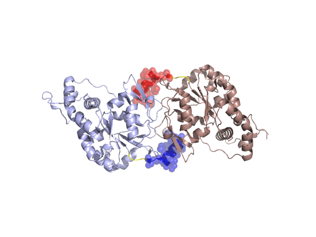
Structural Details of PDB entry 1T8T
Structural Details of PDB entry 1T8T
PDBid Chains Hinge Swapped Domain
1T8T
A,B
A:150-153,B:150-153
A:136-149,B:136-149
Swapped-domain interface residues and interactions:
Chains Residues
A
136 , 137 , 139 , 140 , 141 , 143 , 144 , 147 , 361 , 362 , 363 , 365 ,
B
136 , 137 , 138 , 139 , 140 , 141 , 143 , 144 , 146 , 147 , 361 , 362 , 363 , 365 ,
Non-swapped-domain interface residues and interactions:
Chains Residues
A
174 , 176 , 177 , 179 , 207 , 208 , 210 , 333 , 335 , 337 , 338 , 339 , 340 , 342 , 344 , 345 , 346 , 353 , 354 , 356 , 357 , 358 , 359 , 360 , 366 , 406 ,
B
174 , 176 , 177 , 179 , 207 , 208 , 210 , 333 , 335 , 337 , 338 , 339 , 340 , 342 , 344 , 345 , 353 , 354 , 356 , 357 , 358 , 359 , 360 ,
Mutations in critical regions:
Chains
Hinge
Domain swapped interface Non-swapped interface Swapped Domain
A No mutation PRO(136)-, ASN(137)-, No mutation PRO(136)-, ASN(137)-, SER(138)-, B No mutation PRO(136)-, ASN(137)-, SER(138)-, No mutation PRO(136)-, ASN(137)-, SER(138)-,
HIDE output:
JMOL Visualization:
2D-plot:
JOY Structural annotation for hinge hinge and swapped domain:
JOY output: