Home BrowseSearch Help Group

Pfam Domains mapped on to the structure: 1SJV

No. Chain ID Pfam ID Pfam Description Linkout - Pfam Linkout - CDD
1 A PF13895 Immunoglobulin domain PF13895 PF13895

Conserved Domain Database Superfamily Annotations: 1SJV

No. PDB ID PSSM ID CDD Accession Superfamily Short Name Linkout - CDD
1 1SJV 212623 cl11960 Ig superfamily - -
2 1SJV 212623 cl11960 Ig superfamily - -
Structural Details of PDB entry 1SJV

Structural Details of PDB entry 1SJV

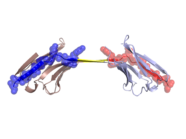
PDBid Chains Hinge Swapped Domain
1SJV A,B A:95-98,B:95-98 A:99-110,B:99-110

Swapped-domain interface residues and interactions:

Chains Residues
A 8, 9, 10, 11, 12, 13, 14, 81, 83, 86, 87, 88, 89, 90, 91, 92, 93, 94, 99, 100, 101, 103, 104, 105, 106, 107, 108, 109, 110,
B 8, 9, 10, 11, 12, 13, 14, 81, 83, 86, 87, 88, 89, 90, 91, 92, 93, 94, 99, 100, 101, 103, 104, 105, 106, 107, 108, 109,

Non-swapped-domain interface residues and interactions:

Chains Residues
A 18, 20, 95, 96, 97, 98,
B 18, 20, 95, 96, 97, 98,

Swapped domains are represented using trasperent spheres.
Non-swapped part is represented using light color and cartoon representation.
Hinge region is shown in yellow color.

Mutations in critical regions:

Chains Hinge Domain swapped interface Non-swapped interface Swapped Domain
ANo mutationNo mutationNo mutationNo mutation
BNo mutationNo mutationNo mutationNo mutation

HIDE output:

Homologues found through HIDE algorithm

JMOL Visualization:

Jmol_logo            Jmol_logo            Jmol_logo

2D-plot:

A:1SJV B:1SJV

JOY Structural annotation for hinge hinge and swapped domain:

Hinge Swapped domain

JOY output:

ali file:1SJV.ali
atm file:1SJV.atm
cof file:1SJV.cof
hbd file:1SJV.hbd
html file:1SJV.html
pdb file:1SJV.pdb
ps file:1SJV.ps
psa file:1SJV.psa
rtf file:1SJV.rtf
sst file:1SJV.sst
tem file:1SJV.tem