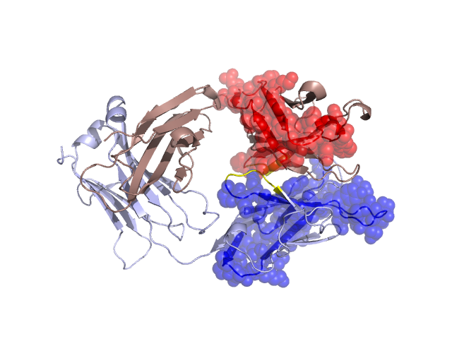
Pfam Domains mapped on to the structure: 1SBS
No.
Chain ID
Pfam ID
Pfam Description
Linkout - Pfam
Linkout - CDD
1
L
PF13895
Immunoglobulin domain
PF13895
PF13895
Conserved Domain Database Superfamily Annotations: 1SBS
Structural Details of PDB entry 1SBS
Structural Details of PDB entry 1SBS
PDBid Chains Hinge Swapped Domain
1SBS
H,L
H:42-46,L:47-51
H:1-41,L:1-46
Swapped-domain interface residues and interactions:
Non-swapped-domain interface residues and interactions:
Chains Residues
H
44 , 45 , 47 , 50 , 52 , 61 , 63 , 64 , 97 , 104 , 106 , 107 , 108 , 109 , 110 , 111 , 113 , 114 , 132 , 133 , 134 , 135 , 136 , 137 , 141 , 143 , 147 , 149 , 151 , 153 , 174 , 176 , 177 , 179 , 181 , 188 , 190 , 192 , 218 , 222 ,
L
49 , 50 , 52 , 55 , 56 , 61 , 93 , 95 , 97 , 100 , 101 , 102 , 104 , 120 , 122 , 124 , 125 , 127 , 129 , 130 , 133 , 137 , 139 , 141 , 143 , 144 , 166 , 168 , 169 , 170 , 173 , 180 , 182 , 184 , 186 , 213 , 219 ,
Mutations in critical regions:
Chains
Hinge
Domain swapped interface Non-swapped interface Swapped Domain
H LYS(43)ARG, No mutation ASP(50)GLU, LEU(61)HIS, TYR(106)-, ASP(107)-, ALA(109)PRO, MET(110)PHE, ASP(111)ALA, ASN(3)LYS, LEU(20)VAL, THR(28)ALA, ASN(31)TYR, L No mutation No mutation GLN(95)LYS, TYR(97)-, -(101)PRO, SER(5)THR, VAL(15)ALA, THR(22)SER, TYR(31)ASN, SER(32)THR, SER(33)TRP, ASN(34)THR, GLN(35)ARG, MET(36)LYS,
HIDE output:
JMOL Visualization:
2D-plot:
JOY Structural annotation for hinge hinge and swapped domain:
JOY output: