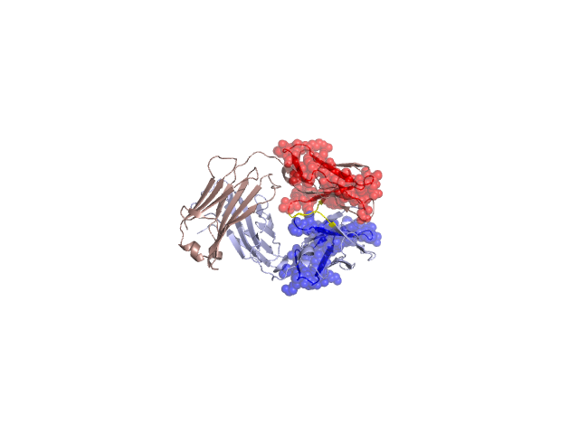
Structural Details of PDB entry 1S5H
Structural Details of PDB entry 1S5H
PDBid Chains Hinge Swapped Domain
1S5H
A,B
A:41-45,B:42-46
A:1-40,B:1-41
Swapped-domain interface residues and interactions:
Non-swapped-domain interface residues and interactions:
Chains Residues
A
42 , 43 , 44 , 46 , 49 , 50 , 87 , 89 , 91 , 94 , 95 , 96 , 98 , 100 , 114 , 116 , 117 , 118 , 119 , 120 , 121 , 123 , 124 , 127 , 131 , 133 , 135 , 137 , 138 , 160 , 161 , 162 , 164 , 167 , 174 , 176 , 178 , 180 , 209 , 211 ,
B
44 , 45 , 47 , 50 , 59 , 61 , 62 , 63 , 95 , 99 , 102 , 103 , 105 , 106 , 108 , 109 , 110 , 127 , 128 , 129 , 130 , 131 , 132 , 134 , 136 , 142 , 146 , 148 , 169 , 171 , 172 , 174 , 176 , 181 , 183 , 185 , 213 , 218 ,