Pfam Domains mapped on to the structure: 1S2W
No.
Chain ID
Pfam ID
Pfam Description
Linkout - Pfam
Linkout - CDD
1
A
PF13714
Phosphoenolpyruvate phosphomutase
PF13714
PF13714
Conserved Domain Database Superfamily Annotations: 1S2W
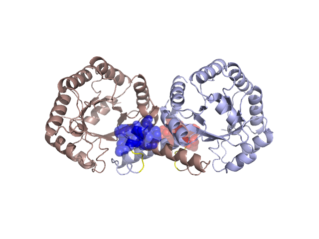
Structural Details of PDB entry 1S2W
Structural Details of PDB entry 1S2W
PDBid Chains Hinge Swapped Domain
1S2W
A,B
A:268-271,B:268-271
A:272-278,B:272-278
Swapped-domain interface residues and interactions:
Non-swapped-domain interface residues and interactions:
Chains Residues
A
22 , 24 , 28 , 29 , 30 , 31 , 33 , 34 , 38 , 39 , 40 , 50 , 51 , 52 , 53 , 54 , 66 , 69 , 73 , 77 , 217 , 218 , 220 , 221 , 224 , 228 , 237 , 239 , 240 , 241 , 242 , 243 , 244 , 245 , 246 , 247 , 248 , 249 , 250 , 252 , 253 , 256 , 257 , 260 , 261 , 262 , 263 , 266 , 268 , 269 , 270 , 271 ,
B
22 , 24 , 28 , 29 , 30 , 31 , 33 , 34 , 38 , 39 , 40 , 51 , 52 , 53 , 54 , 66 , 69 , 73 , 77 , 217 , 218 , 220 , 221 , 224 , 228 , 237 , 239 , 240 , 241 , 242 , 243 , 244 , 245 , 246 , 247 , 248 , 249 , 250 , 252 , 253 , 256 , 257 , 260 , 261 , 262 , 263 , 266 , 268 , 269 , 270 , 271 ,
Mutations in critical regions:
Chains
Hinge
Domain swapped interface Non-swapped interface Swapped Domain
A No mutation No mutation No mutation No mutation B No mutation No mutation No mutation No mutation
HIDE output:
JMOL Visualization:
2D-plot:
JOY Structural annotation for hinge hinge and swapped domain:
JOY output: