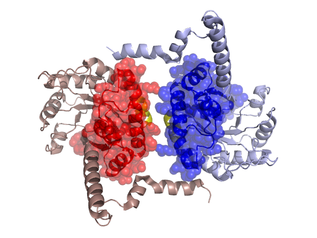
Pfam Domains mapped on to the structure: 1S2U
No.
Chain ID
Pfam ID
Pfam Description
Linkout - Pfam
Linkout - CDD
1
A
PF13714
Phosphoenolpyruvate phosphomutase
PF13714
PF13714
Conserved Domain Database Superfamily Annotations: 1S2U
Structural Details of PDB entry 1S2U
Structural Details of PDB entry 1S2U
PDBid Chains Hinge Swapped Domain
1S2U
A,B
A:100-106,B:100-106
A:5-99,B:5-99
Swapped-domain interface residues and interactions:
Chains Residues
A
65 , 72 , 73 , 76 , 92 , 93 , 94 , 97 , 98 , 291 ,
B
65 , 69 , 72 , 73 , 76 , 92 , 93 , 94 , 97 , 98 , 291 ,
Non-swapped-domain interface residues and interactions:
Chains Residues
A
102 , 105 , 106 , 136 , 137 , 140 , 143 , 144 , 147 , 283 , 284 , 287 , 288 , 290 , 292 , 293 , 294 ,
B
102 , 105 , 106 , 136 , 137 , 140 , 143 , 144 , 147 , 283 , 284 , 287 , 288 , 290 , 292 ,
Mutations in critical regions:
Chains
Hinge
Domain swapped interface Non-swapped interface Swapped Domain
A No mutation No mutation No mutation MET(14)ALA, MET(24)ALA, ALA(58)ASP, MET(74)ALA, B No mutation No mutation No mutation MET(14)ALA, MET(24)ALA, ALA(58)ASP, MET(74)ALA,
HIDE output:
JMOL Visualization:
2D-plot:
JOY Structural annotation for hinge hinge and swapped domain:
JOY output: