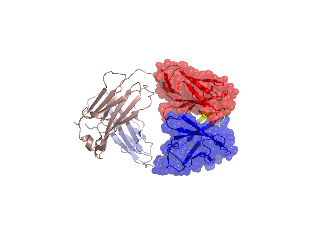
Structural Details of PDB entry 1RJL
Structural Details of PDB entry 1RJL
PDBid Chains Hinge Swapped Domain
1RJL
A,B
A:93-97,B:102-108
A:1-92,B:1-101
Swapped-domain interface residues and interactions:
Chains Residues
A
32 , 34 , 36 , 38 , 43 , 44 , 46 , 49 , 50 , 55 , 56 , 87 , 89 , 91 , 95 , 96 , 98 , 100 ,
B
39 , 44 , 45 , 47 , 50 , 59 , 61 , 62 , 95 , 99 , 104 , 106 , 108 , 112 ,
Non-swapped-domain interface residues and interactions:
Chains Residues
A
94 , 116 , 117 , 118 , 119 , 120 , 121 , 123 , 124 , 127 , 131 , 133 , 135 , 137 , 138 , 160 , 161 , 162 , 163 , 164 , 167 , 174 , 176 , 178 , 180 , 209 , 212 ,
B
103 , 105 , 107 , 110 , 111 , 129 , 130 , 131 , 132 , 133 , 134 , 135 , 144 , 146 , 148 , 150 , 171 , 172 , 173 , 174 , 176 , 178 , 183 , 185 , 187 , 215 , 220 ,