Home BrowseSearch Help Group

Pfam Domains mapped on to the structure: 1R9C

No. Chain ID Pfam ID Pfam Description Linkout - Pfam Linkout - CDD
1 A PF00903 Glyoxalase/Bleomycin resistance protein/Dioxygenase superfamily PF00903 PF00903
2 A PF12681 Glyoxalase-like domain PF12681 PF12681

Conserved Domain Database Superfamily Annotations: 1R9C

No. PDB ID PSSM ID CDD Accession Superfamily Short Name Linkout - CDD
1 1R9C 176712 cd08364 FosX - cl14632
2 1R9C 211465 cl14632 Glo_EDI_BRP_like superfamily - -
3 1R9C 211465 cl14632 Glo_EDI_BRP_like superfamily - -
Structural Details of PDB entry 1R9C

Structural Details of PDB entry 1R9C

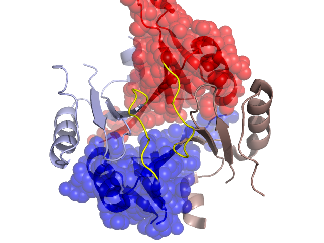
PDBid Chains Hinge Swapped Domain
1R9C A,B A:60-68,B:60-68 A:1-59,B:1-59

Swapped-domain interface residues and interactions:

Chains Residues
A 1, 2, 3, 4, 5, 6, 7, 8, 9, 11, 26, 31, 32, 44, 46, 48, 49, 52, 53, 67, 68, 69, 70, 71, 72, 73, 74, 120,
B 1, 2, 3, 4, 5, 6, 7, 8, 9, 11, 26, 27, 31, 32, 44, 46, 48, 49, 52, 53, 57, 67, 68, 69, 70, 71, 72, 73, 74, 120,

Non-swapped-domain interface residues and interactions:

Chains Residues
A 62, 63, 64, 66, 75, 78, 82, 124, 127, 128, 130,
B 62, 63, 64, 66, 75, 78, 82, 124, 127, 128,

Swapped domains are represented using trasperent spheres.
Non-swapped part is represented using light color and cartoon representation.
Hinge region is shown in yellow color.

Mutations in critical regions:

Chains Hinge Domain swapped interface Non-swapped interface Swapped Domain
ANo mutationNo mutationNo mutationNo mutation
BNo mutationNo mutationNo mutationNo mutation

HIDE output:

Homologues found through HIDE algorithm

JMOL Visualization:

Jmol_logo            Jmol_logo            Jmol_logo

2D-plot:

A:1R9C B:1R9C

JOY Structural annotation for hinge hinge and swapped domain:

Hinge Swapped domain

JOY output:

ali file:1R9C.ali
atm file:1R9C.atm
cof file:1R9C.cof
hbd file:1R9C.hbd
html file:1R9C.html
pdb file:1R9C.pdb
ps file:1R9C.ps
psa file:1R9C.psa
rtf file:1R9C.rtf
sst file:1R9C.sst
tem file:1R9C.tem