Home BrowseSearch Help Group

Pfam Domains mapped on to the structure: 1R7H

No. Chain ID Pfam ID Pfam Description Linkout - Pfam Linkout - CDD
1 A PF00462 Glutaredoxin PF00462 PF00462

Gene Ontology Annotations: 1R7H

No. GO ID GO Description Linkout - AmiGO
1 GO:0045454 cell redox homeostasis GO:0045454

Conserved Domain Database Superfamily Annotations: 1R7H

No. PDB ID PSSM ID CDD Accession Superfamily Short Name Linkout - CDD
1 1R7H 48525 cd02976 NrdH - cl00388
2 1R7H 212209 cl00388 Thioredoxin_like superfamily - -
Structural Details of PDB entry 1R7H

Structural Details of PDB entry 1R7H

PDBid Chains Hinge Swapped Domain
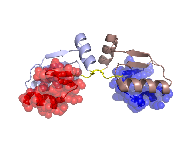
1R7H A,B A:47-52,B:47-52 A:53-74,B:53-74

Swapped-domain interface residues and interactions:

Chains Residues
A 1, 2, 3, 4, 5, 6, 13, 14, 16, 17, 21, 24, 53, 54, 55, 56, 57, 60, 62, 63, 64, 69, 72, 73, 74,
B 1, 2, 3, 4, 5, 6, 13, 14, 16, 17, 21, 24, 53, 54, 55, 56, 57, 60, 62, 63, 64, 69, 72, 73,

Non-swapped-domain interface residues and interactions:

Chains Residues
A 8, 11, 20, 26, 33, 34, 37, 40, 41, 42, 43, 44, 46, 47, 48, 49, 50, 51, 52,
B 8, 11, 20, 26, 33, 37, 40, 41, 42, 43, 44, 46, 47, 48, 49, 50, 51, 52,

Swapped domains are represented using trasperent spheres.
Non-swapped part is represented using light color and cartoon representation.
Hinge region is shown in yellow color.

Mutations in critical regions:

Chains Hinge Domain swapped interface Non-swapped interface Swapped Domain
ANo mutationNo mutationNo mutationNo mutation
BNo mutationNo mutationNo mutationNo mutation

HIDE output:

Homologues found through HIDE algorithm

JMOL Visualization:

Jmol_logo            Jmol_logo            Jmol_logo

2D-plot:

A:1R7H B:1R7H

JOY Structural annotation for hinge hinge and swapped domain:

Hinge Swapped domain

JOY output:

ali file:1R7H.ali
atm file:1R7H.atm
cof file:1R7H.cof
hbd file:1R7H.hbd
html file:1R7H.html
pdb file:1R7H.pdb
ps file:1R7H.ps
psa file:1R7H.psa
rtf file:1R7H.rtf
sst file:1R7H.sst
tem file:1R7H.tem