Home BrowseSearch Help Group

Pfam Domains mapped on to the structure: 1R5D

No. Chain ID Pfam ID Pfam Description Linkout - Pfam Linkout - CDD
1 A PF00074 Pancreatic ribonuclease PF00074 PF00074

Conserved Domain Database Superfamily Annotations: 1R5D

No. PDB ID PSSM ID CDD Accession Superfamily Short Name Linkout - CDD
1 1R5D 119387 cd06265 RNase_A_canonical - cl00128
2 1R5D 206850 cl00128 RNase_A superfamily - -
3 1R5D 206850 cl00128 RNase_A superfamily - -
Structural Details of PDB entry 1R5D

Structural Details of PDB entry 1R5D

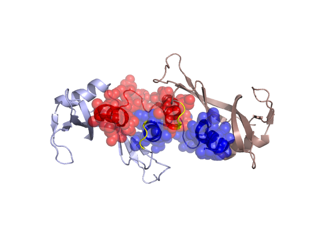
PDBid Chains Hinge Swapped Domain
1R5D A,B A:33-35,B:33-35 A:1-32,B:1-32

Swapped-domain interface residues and interactions:

Chains Residues
A 4, 5, 8, 9, 10, 11, 12, 13, 14, 15, 16, 17, 19, 20, 22, 25, 28, 29, 31, 32, 33, 45, 46, 47, 48, 49, 50, 116, 117, 118,
B 4, 5, 8, 9, 10, 11, 12, 13, 14, 15, 17, 18, 19, 20, 22, 25, 28, 29, 31, 32, 33, 45, 46, 47, 48, 49, 50, 116, 117, 118,

Non-swapped-domain interface residues and interactions:

Chains Residues
A 34, 35, 41, 44, 51, 54, 80, 101, 120, 124,
B 34, 35, 37, 41, 44, 51, 54, 80, 101, 120,

Swapped domains are represented using trasperent spheres.
Non-swapped part is represented using light color and cartoon representation.
Hinge region is shown in yellow color.

Mutations in critical regions:

Chains Hinge Domain swapped interface Non-swapped interface Swapped Domain
ANo mutationNo mutationNo mutationNo mutation
BNo mutationNo mutationNo mutationNo mutation

HIDE output:

Homologues found through HIDE algorithm

JMOL Visualization:

Jmol_logo            Jmol_logo            Jmol_logo

2D-plot:

A:1R5D B:1R5D

JOY Structural annotation for hinge hinge and swapped domain:

Hinge Swapped domain

JOY output:

ali file:1R5D.ali
atm file:1R5D.atm
cof file:1R5D.cof
hbd file:1R5D.hbd
html file:1R5D.html
pdb file:1R5D.pdb
ps file:1R5D.ps
psa file:1R5D.psa
rtf file:1R5D.rtf
sst file:1R5D.sst
tem file:1R5D.tem