Home BrowseSearch Help Group

Pfam Domains mapped on to the structure: 1R1T

No. Chain ID Pfam ID Pfam Description Linkout - Pfam Linkout - CDD
1 A PF12840 Helix-turn-helix domain PF12840 PF12840
2 A PF01022 Bacterial regulatory protein, arsR family PF01022 PF01022

Conserved Domain Database Superfamily Annotations: 1R1T

No. PDB ID PSSM ID CDD Accession Superfamily Short Name Linkout - CDD
1 1R1T 28974 cd00090 HTH_ARSR - cl00088
2 1R1T 212173 cl00088 HTH superfamily - -
Structural Details of PDB entry 1R1T

Structural Details of PDB entry 1R1T

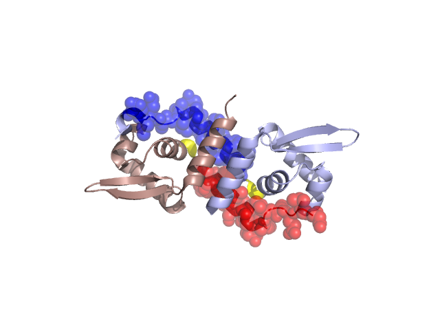
PDBid Chains Hinge Swapped Domain
1R1T A,B A:44-46,B:44-46 A:24-43,B:24-43

Swapped-domain interface residues and interactions:

Chains Residues
A 25, 26, 27, 28, 30, 32, 33, 34, 35, 36, 37, 39, 40, 41, 43, 44, 46, 49, 50, 53, 54, 68, 115,
B 25, 26, 27, 28, 30, 32, 33, 34, 35, 36, 37, 39, 40, 41, 43, 44, 46, 49, 50, 53, 54, 68, 115,

Non-swapped-domain interface residues and interactions:

Chains Residues
A 57, 58, 60, 64, 87, 104, 106, 107, 109, 110, 111, 113, 114, 116, 117, 118, 121,
B 20, 21, 22, 57, 58, 60, 87, 103, 106, 107, 109, 110, 111, 113, 114, 116, 117,

Swapped domains are represented using trasperent spheres.
Non-swapped part is represented using light color and cartoon representation.
Hinge region is shown in yellow color.

Mutations in critical regions:

Chains Hinge Domain swapped interface Non-swapped interface Swapped Domain
ANo mutationNo mutationNo mutationNo mutation
BNo mutationNo mutationNo mutationNo mutation

HIDE output:

Homologues found through HIDE algorithm

JMOL Visualization:

Jmol_logo            Jmol_logo            Jmol_logo

2D-plot:

A:1R1T B:1R1T

JOY Structural annotation for hinge hinge and swapped domain:

Hinge Swapped domain

JOY output:

ali file:1R1T.ali
atm file:1R1T.atm
cof file:1R1T.cof
hbd file:1R1T.hbd
html file:1R1T.html
pdb file:1R1T.pdb
ps file:1R1T.ps
psa file:1R1T.psa
rtf file:1R1T.rtf
sst file:1R1T.sst
tem file:1R1T.tem