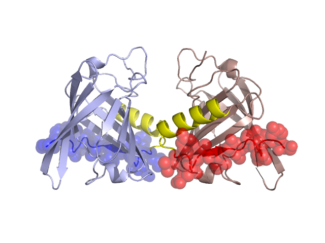
Pfam Domains mapped on to the structure: 1QY2 No. Chain ID Pfam ID Pfam Description Linkout - Pfam Linkout - CDD 1 A PF00061 Lipocalin / cytosolic fatty-acid binding protein family PF00061 PF00061 Conserved Domain Database Superfamily Annotations: 1QY2 No. PDB ID PSSM ID CDD Accession Superfamily Short Name Linkout - CDD 1 1QY2 207329 cl01150 Lipocalin superfamily - - Structural Details of PDB entry 1QY2 Structural Details of PDB entry 1QY2 PDBid Chains Hinge Swapped Domain 1QY2 A,B A:128-143,B:128-143 A:144-157,B:144-157 Swapped-domain interface residues and interactions: Chains Residues A 144, 157, B 144, Non-swapped-domain interface residues and interactions: Chains Residues A 27, 76, 79, 104, 106, 108, 111, 112, 113, 115, 139, 140, 141, 142, B 27, 76, 79, 104, 106, 108, 111, 112, 113, 115, 139, 140, 141, 142, Swapped domains are represented using trasperent spheres. Non-swapped part is represented using light color and cartoon representation. Hinge region is shown in yellow color. Mutations in critical regions: Chains Hinge Domain swapped interface Non-swapped interface Swapped Domain ANo mutationNo mutationNo mutationNo mutation BNo mutationNo mutationNo mutationNo mutation HIDE output: Homologues found through HIDE algorithm JMOL Visualization: 2D-plot: A:1QY2 B:1QY2 JOY Structural annotation for hinge hinge and swapped domain: Hinge Swapped domain JOY output: ali file:1QY2.ali atm file:1QY2.atm cof file:1QY2.cof hbd file:1QY2.hbd html file:1QY2.html pdb file:1QY2.pdb ps file:1QY2.ps psa file:1QY2.psa rtf file:1QY2.rtf sst file:1QY2.sst tem file:1QY2.tem