Pfam Domains mapped on to the structure: 1QB2
No.
Chain ID
Pfam ID
Pfam Description
Linkout - Pfam
Linkout - CDD
1
A
PF02978
Signal peptide binding domain
PF02978
PF02978
Gene Ontology Annotations: 1QB2
Conserved Domain Database Superfamily Annotations: 1QB2
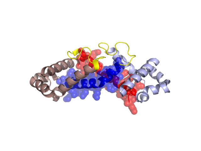
Structural Details of PDB entry 1QB2
Structural Details of PDB entry 1QB2
PDBid Chains Hinge Swapped Domain
1QB2
A,B
A:351-365,B:351-365
A:326-350,B:326-350
Swapped-domain interface residues and interactions:
Chains Residues
A
326 , 327 , 328 , 329 , 330 , 332 , 333 , 335 , 336 , 337 , 338 , 339 , 340 , 341 , 342 , 343 , 345 , 346 , 347 , 349 , 362 , 365 , 369 , 372 , 376 , 387 , 388 , 389 , 390 , 392 , 428 ,
B
326 , 327 , 328 , 329 , 330 , 332 , 333 , 335 , 336 , 337 , 338 , 339 , 340 , 342 , 343 , 344 , 345 , 346 , 347 , 349 , 350 , 369 , 372 , 376 , 387 , 388 , 389 , 390 , 392 , 428 ,
Non-swapped-domain interface residues and interactions:
Chains Residues
A
358 , 360 , 361 , 366 , 373 , 375 , 379 , 380 , 384 , 420 , 424 , 427 , 431 ,
B
359 , 360 , 361 , 362 , 365 , 373 , 375 , 379 , 380 , 382 , 384 , 420 , 424 , 427 , 431 ,
Mutations in critical regions:
Chains
Hinge
Domain swapped interface Non-swapped interface Swapped Domain
A No mutation No mutation No mutation No mutation B No mutation No mutation No mutation No mutation
HIDE output:
JMOL Visualization:
2D-plot:
JOY Structural annotation for hinge hinge and swapped domain:
JOY output: