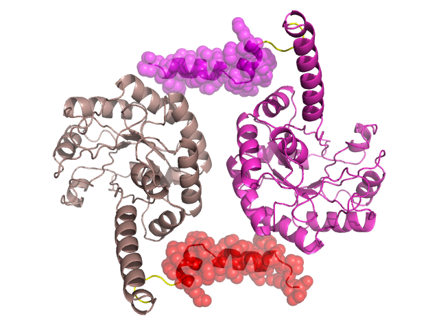
Structural Details of PDB entry 1PYM
Structural Details of PDB entry 1PYM
PDBid Chains Hinge Swapped Domain
1PYM
A,D,C,B
A:267-271,D:267-271,C:267-271,B:267-271
A:272-295,D:272-295,C:272-295,B:272-295
Swapped-domain interface residues and interactions:
Chains Residues
A
93 , 137 , 140 , 144 , 283 , 284 , 287 , 288 , 290 , 291 , 292 , 293 , 295 ,
B
93 , 136 , 137 , 140 , 144 , 283 , 284 , 287 , 290 , 291 , 292 ,
Non-swapped-domain interface residues and interactions:
Chains Residues
A
65 , 72 , 73 , 76 , 92 , 94 , 97 , 98 , 101 , 102 , 105 , 106 , 136 , 147 ,
B
65 , 72 , 73 , 76 , 92 , 94 , 97 , 98 , 101 , 102 , 105 , 106 , 143 , 147 ,