Home BrowseSearch Help Group

Pfam Domains mapped on to the structure: 1OI6

No. Chain ID Pfam ID Pfam Description Linkout - Pfam Linkout - CDD
1 A PF00908 dTDP-4-dehydrorhamnose 3,5-epimerase PF00908 PF00908

Gene Ontology Annotations: 1OI6

No. GO ID GO Description Linkout - AmiGO
1 GO:0009103 lipopolysaccharide biosynthetic process GO:0009103

Conserved Domain Database Superfamily Annotations: 1OI6

No. PDB ID PSSM ID CDD Accession Superfamily Short Name Linkout - CDD
1 1OI6 212615 cl09118 Cupin_2 superfamily - -
Structural Details of PDB entry 1OI6

Structural Details of PDB entry 1OI6

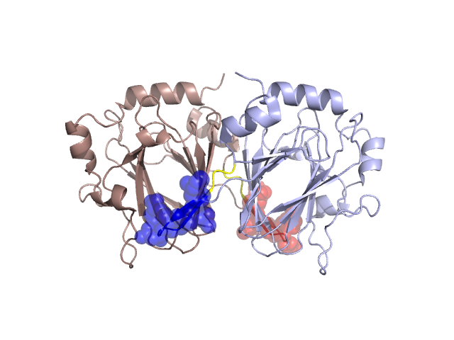
PDBid Chains Hinge Swapped Domain
1OI6 A,B A:16-18,B:16-18;A:27-29,B:27-29 A:19-26,B:19-26

Swapped-domain interface residues and interactions:

Chains Residues
A 22, 23, 24, 25, 26, 51, 52, 53, 55, 58, 165,
B 22, 23, 24, 25, 26, 51, 52, 53, 55, 58, 165,

Non-swapped-domain interface residues and interactions:

Chains Residues
A 27, 28, 29, 30, 31, 32, 42, 43, 45, 46, 47, 48, 49, 50, 60, 76, 78, 79, 106, 132, 134, 138, 166, 167, 200, 202,
B 27, 28, 29, 30, 31, 32, 42, 43, 45, 46, 47, 48, 49, 50, 60, 76, 78, 79, 106, 132, 134, 138, 167, 200, 202,

Swapped domains are represented using trasperent spheres.
Non-swapped part is represented using light color and cartoon representation.
Hinge region is shown in yellow color.

Mutations in critical regions:

Chains Hinge Domain swapped interface Non-swapped interface Swapped Domain
ANo mutationNo mutationNo mutationNo mutation
BNo mutationNo mutationNo mutationNo mutation

HIDE output:

Homologues found through HIDE algorithm

JMOL Visualization:

Jmol_logo            Jmol_logo            Jmol_logo

2D-plot:

A:1OI6 B:1OI6

JOY Structural annotation for hinge hinge and swapped domain:

Hinge Swapped domain

JOY output:

ali file:1OI6.ali
atm file:1OI6.atm
cof file:1OI6.cof
hbd file:1OI6.hbd
html file:1OI6.html
pdb file:1OI6.pdb
ps file:1OI6.ps
psa file:1OI6.psa
rtf file:1OI6.rtf
sst file:1OI6.sst
tem file:1OI6.tem