Pfam Domains mapped on to the structure: 1OAS
No.
Chain ID
Pfam ID
Pfam Description
Linkout - Pfam
Linkout - CDD
1
A
PF00291
Pyridoxal-phosphate dependent enzyme
PF00291
PF00291
Conserved Domain Database Superfamily Annotations: 1OAS
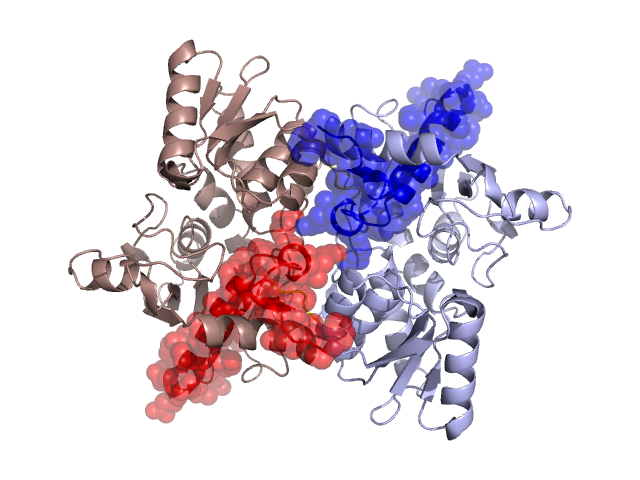
Structural Details of PDB entry 1OAS
Structural Details of PDB entry 1OAS
PDBid Chains Hinge Swapped Domain
1OAS
A,B
A:266-269,B:266-269
A:270-315,B:270-315
Swapped-domain interface residues and interactions:
Chains Residues
A
98 , 101 , 104 , 301 , 303 , 304 , 306 , 307 , 311 , 314 , 315 ,
B
98 , 101 , 104 , 301 , 303 , 304 , 306 , 307 , 311 , 314 ,
Non-swapped-domain interface residues and interactions:
Chains Residues
A
1 , 2 , 3 , 4 , 5 , 6 , 7 , 10 , 12 , 15 , 16 , 17 , 18 , 20 , 21 , 28 , 33 , 34 , 35 , 36 , 37 , 38 , 78 , 81 , 82 , 97 , 100 , 102 , 105 , 106 , 107 , 163 , 164 , 165 , 262 , 263 , 265 , 266 , 267 , 268 ,
B
1 , 2 , 3 , 4 , 5 , 6 , 7 , 10 , 12 , 15 , 16 , 17 , 18 , 20 , 28 , 33 , 34 , 35 , 36 , 37 , 38 , 78 , 81 , 82 , 84 , 97 , 102 , 105 , 106 , 107 , 163 , 164 , 165 , 262 , 263 , 264 , 265 , 266 , 267 , 268 ,
Mutations in critical regions:
Chains
Hinge
Domain swapped interface Non-swapped interface Swapped Domain
A GLY(266)VAL, ILE(267)PHE, No mutation GLY(266)VAL, ILE(267)PHE, No mutation B GLY(266)VAL, ILE(267)PHE, No mutation GLY(266)VAL, ILE(267)PHE, No mutation
HIDE output:
JMOL Visualization:
2D-plot:
JOY Structural annotation for hinge hinge and swapped domain:
JOY output: