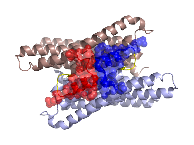
Structural Details of PDB entry 1O5H
Structural Details of PDB entry 1O5H
PDBid Chains Hinge Swapped Domain
1O5H
A,B
A:19-23,B:19-23
A:2-18,B:2-18
Swapped-domain interface residues and interactions:
Chains Residues
A
4 , 5 , 6 , 7 , 8 , 9 , 12 , 13 , 15 , 16 , 18 , 37 , 38 , 40 , 44 , 45 ,
B
4 , 5 , 6 , 7 , 8 , 9 , 12 , 13 , 15 , 16 , 18 , 37 , 38 , 40 , 44 , 45 ,
Non-swapped-domain interface residues and interactions:
Chains Residues
A
19 , 20 , 23 , 24 , 27 , 30 , 31 , 33 , 48 , 70 , 74 , 136 , 137 , 139 , 140 , 143 , 144 , 147 , 150 , 151 , 154 , 155 , 157 , 158 , 161 , 191 , 197 , 198 , 199 , 200 , 201 ,
B
19 , 20 , 23 , 24 , 27 , 30 , 31 , 33 , 48 , 70 , 74 , 136 , 137 , 139 , 140 , 143 , 144 , 147 , 150 , 151 , 154 , 155 , 157 , 158 , 161 , 162 , 191 , 197 , 198 ,