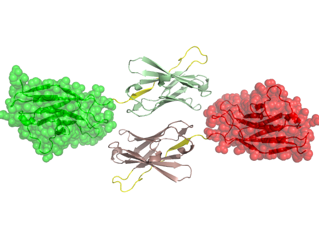
Pfam Domains mapped on to the structure: 1NQB No. Chain ID Pfam ID Pfam Description Linkout - Pfam Linkout - CDD 1 A PF13895 Immunoglobulin domain PF13895 PF13895 2 A PF13895 Immunoglobulin domain PF13895 PF13895 Conserved Domain Database Superfamily Annotations: 1NQB No. PDB ID PSSM ID CDD Accession Superfamily Short Name Linkout - CDD 1 1NQB 143182 cd04981 IgV_H - cl11960 2 1NQB 212623 cl11960 Ig superfamily - - 3 1NQB 143181 cd04980 IgV_L_kappa - cl11960 4 1NQB 212623 cl11960 Ig superfamily - - 5 1NQB 212623 cl11960 Ig superfamily - - 6 1NQB 212623 cl11960 Ig superfamily - - Structural Details of PDB entry 1NQB Structural Details of PDB entry 1NQB PDBid Chains Hinge Swapped Domain 1NQB F,A,D,C,E,B F:99-120,A:99-120,D:99-120,C:99-120,E:99-120,B:99-120 F:121-233,A:121-233,D:121-233,C:121-233,E:121-233,B:121-233 Swapped-domain interface residues and interactions: Chains Residues A 151, 152, 233, C 151, 152, Non-swapped-domain interface residues and interactions: Chains Residues A 16, 17, 19, 55, 74, 82, C 17, 19, 54, 55, 74, 82, Swapped domains are represented using trasperent spheres. Non-swapped part is represented using light color and cartoon representation. Hinge region is shown in yellow color. Mutations in critical regions: Chains Hinge Domain swapped interface Non-swapped interface Swapped Domain FNo mutationNo mutationNo mutationNo mutation ANo mutationNo mutationNo mutationNo mutation DNo mutationNo mutationNo mutationNo mutation CNo mutationNo mutationNo mutationNo mutation ENo mutationNo mutationNo mutationNo mutation BNo mutationNo mutationNo mutationNo mutation HIDE output: Homologues found through HIDE algorithm JMOL Visualization: 2D-plot: A:1NQB B:1NQB C:1NQB D:1NQB E:1NQB F:1NQB JOY Structural annotation for hinge hinge and swapped domain: Hinge Swapped domain JOY output: ali file:1NQB.ali atm file:1NQB.atm cof file:1NQB.cof hbd file:1NQB.hbd html file:1NQB.html pdb file:1NQB.pdb ps file:1NQB.ps psa file:1NQB.psa rtf file:1NQB.rtf sst file:1NQB.sst tem file:1NQB.tem