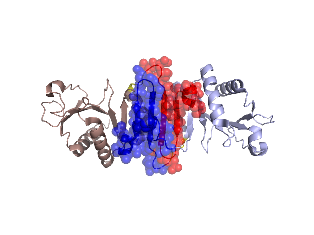
Structural Details of PDB entry 1NP6
Structural Details of PDB entry 1NP6
PDBid Chains Hinge Swapped Domain
1NP6
A,B
A:86-89,B:86-89
A:47-85,B:47-85
Swapped-domain interface residues and interactions:
Chains Residues
A
25 , 28 , 29 , 32 , 38 , 39 , 40 , 41 , 42 , 43 , 44 , 46 , 59 , 60 , 61 , 63 , 64 , 65 , 66 , 67 , 68 , 69 , 70 , 71 , 72 , 73 , 74 , 75 , 76 , 77 , 78 , 79 , 80 , 81 , 82 , 86 ,
B
25 , 28 , 29 , 32 , 38 , 39 , 40 , 41 , 42 , 43 , 44 , 47 , 48 , 49 , 53 , 56 , 59 , 60 , 63 , 64 , 65 , 66 , 67 , 68 , 69 , 70 , 71 , 72 , 73 , 74 , 75 , 76 , 77 , 78 , 79 , 80 , 81 , 82 , 86 ,
Non-swapped-domain interface residues and interactions:
Chains Residues
A
87 , 92 , 93 , 96 , 97 , 98 , 101 , 102 , 175 ,
B
21 , 46 , 87 , 92 , 93 , 96 , 97 , 98 , 101 , 102 ,