Pfam Domains mapped on to the structure: 1N2F
No.
Chain ID
Pfam ID
Pfam Description
Linkout - Pfam
Linkout - CDD
1
A
PF02566
OsmC-like protein
PF02566
PF02566
Conserved Domain Database Superfamily Annotations: 1N2F
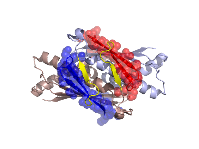
Structural Details of PDB entry 1N2F
Structural Details of PDB entry 1N2F
PDBid Chains Hinge Swapped Domain
1N2F
A,B
A:25-33,B:25-33
A:1-24,B:1-24
Swapped-domain interface residues and interactions:
Chains Residues
A
1 , 2 , 3 , 4 , 6 , 7 , 8 , 9 , 10 , 11 , 12 , 13 , 14 , 15 , 16 , 17 , 18 , 22 , 24 , 38 , 39 , 50 , 51 , 54 , 55 , 58 , 59 , 60 , 62 , 63 , 80 , 81 , 82 , 83 , 84 , 85 , 86 , 87 , 88 , 89 , 90 , 91 , 96 , 97 , 98 , 132 , 133 , 134 , 135 ,
B
1 , 2 , 3 , 4 , 5 , 6 , 7 , 8 , 9 , 10 , 11 , 12 , 13 , 14 , 15 , 16 , 17 , 18 , 22 , 24 , 38 , 39 , 50 , 51 , 54 , 55 , 58 , 59 , 60 , 62 , 63 , 80 , 81 , 82 , 83 , 84 , 85 , 86 , 87 , 88 , 89 , 90 , 91 , 96 , 98 , 132 , 133 , 134 , 135 ,
Non-swapped-domain interface residues and interactions:
Chains Residues
A
26 , 28 , 29 , 31 , 36 , 45 , 46 , 47 , 48 , 49 , 52 , 53 , 56 , 66 , 67 , 92 , 93 , 94 , 95 , 122 , 123 , 125 , 126 , 128 , 129 , 130 , 131 , 142 ,
B
26 , 28 , 29 , 31 , 36 , 45 , 46 , 47 , 48 , 49 , 52 , 53 , 56 , 66 , 67 , 92 , 93 , 94 , 95 , 97 , 122 , 123 , 125 , 126 , 128 , 129 , 130 , 131 ,
Mutations in critical regions:
Chains
Hinge
Domain swapped interface Non-swapped interface Swapped Domain
A No mutation MET(39)LEU, No mutation No mutation B No mutation MET(39)LEU, No mutation No mutation
HIDE output:
JMOL Visualization:
2D-plot:
JOY Structural annotation for hinge hinge and swapped domain:
JOY output: