Structural Details of PDB entry 1MW5
Structural Details of PDB entry 1MW5
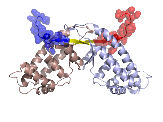
PDBid Chains Hinge Swapped Domain
1MW5
A,B
A:147-150,B:147-150
A:151-164,B:151-164
Swapped-domain interface residues and interactions:
Chains Residues
A
102 , 105 , 142 , 143 , 144 , 145 , 152 , 153 , 154 , 157 , 158 , 159 , 164 ,
B
102 , 105 , 142 , 143 , 144 , 145 , 152 , 153 , 154 , 157 , 158 , 159 ,
Non-swapped-domain interface residues and interactions:
Chains Residues
A
3 , 7 , 11 , 14 , 15 , 16 , 17 , 99 , 103 , 106 , 109 , 114 , 116 , 117 , 119 , 122 , 124 , 126 , 129 , 138 , 146 , 147 , 148 , 149 , 150 ,
B
3 , 7 , 11 , 14 , 15 , 16 , 17 , 99 , 103 , 106 , 109 , 114 , 116 , 117 , 119 , 122 , 124 , 126 , 129 , 138 , 146 , 147 , 148 , 149 , 150 ,
Mutations in critical regions:
Chains
Hinge
Domain swapped interface Non-swapped interface Swapped Domain
A No mutation LEU(157)-, GLU(158)-, TYR(159)-, ASN(164)-, No mutation LEU(157)-, GLU(158)-, TYR(159)-, LYS(160)-, GLY(161)-, GLU(162)-, LEU(163)-, ASN(164)-, B No mutation LEU(157)-, GLU(158)-, TYR(159)-, No mutation LEU(157)-, GLU(158)-, TYR(159)-, LYS(160)-, GLY(161)-, GLU(162)-, LEU(163)-, ASN(164)-,
HIDE output:
JMOL Visualization:
2D-plot:
JOY Structural annotation for hinge hinge and swapped domain:
JOY output: