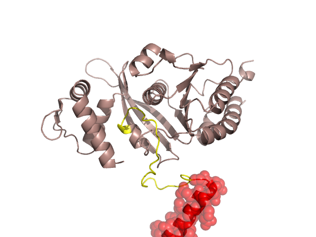
Pfam Domains mapped on to the structure: 1MO6 No. Chain ID Pfam ID Pfam Description Linkout - Pfam Linkout - CDD 1 A PF00154 recA bacterial DNA recombination protein PF00154 PF00154 2 A PF08423 Rad51 PF08423 PF08423 3 A PF06745 KaiC PF06745 PF06745 Conserved Domain Database Superfamily Annotations: 1MO6 No. PDB ID PSSM ID CDD Accession Superfamily Short Name Linkout - CDD 1 1MO6 29984 cd00983 recA - cl09099 2 1MO6 212613 cl09099 P-loop_NTPase superfamily - - 3 1MO6 212613 cl09099 P-loop_NTPase superfamily - - Structural Details of PDB entry 1MO6 Structural Details of PDB entry 1MO6 PDBid Chains Hinge Swapped Domain 1MO6 A,B A:24-47,B:24-47 A:1-23,B:1-23 Swapped-domain interface residues and interactions: Chains Residues Non-swapped-domain interface residues and interactions: Chains Residues Swapped domains are represented using trasperent spheres. Non-swapped part is represented using light color and cartoon representation. Hinge region is shown in yellow color. Mutations in critical regions: Chains Hinge Domain swapped interface Non-swapped interface Swapped Domain ANo mutationNo mutationNo mutationNo mutation BNo mutationNo mutationNo mutationNo mutation HIDE output: Homologues found through HIDE algorithm JMOL Visualization: 2D-plot: A:1MO6 JOY Structural annotation for hinge hinge and swapped domain: Hinge Swapped domain JOY output: ali file:1MO6.ali atm file:1MO6.atm cof file:1MO6.cof hbd file:1MO6.hbd html file:1MO6.html pdb file:1MO6.pdb ps file:1MO6.ps psa file:1MO6.psa rtf file:1MO6.rtf sst file:1MO6.sst tem file:1MO6.tem