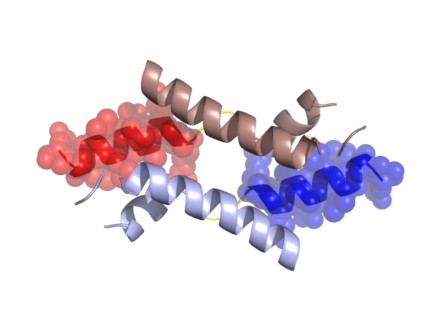
Pfam Domains mapped on to the structure: 1MN3 No. Chain ID Pfam ID Pfam Description Linkout - Pfam Linkout - CDD 1 A PF02845 CUE domain PF02845 PF02845 Gene Ontology Annotations: 1MN3 No. GO ID GO Description Linkout - AmiGO 1 GO:0005515 protein binding GO:0005515 Conserved Domain Database Superfamily Annotations: 1MN3 No. PDB ID PSSM ID CDD Accession Superfamily Short Name Linkout - CDD 1 1MN3 207689 cl02655 CUE superfamily - - Structural Details of PDB entry 1MN3 Structural Details of PDB entry 1MN3 PDBid Chains Hinge Swapped Domain 1MN3 A,B A:434-437,B:434-437 A:438-451,B:438-451 Swapped-domain interface residues and interactions: Chains Residues A 431, 432, 438, 439, 442, 443, 445, 446, 447, 449, 450, 451, B 431, 432, 438, 439, 442, 443, 445, 446, 447, 449, 450, Non-swapped-domain interface residues and interactions: Chains Residues A 404, 412, 415, 416, 420, 427, 428, 430, 434, 435, B 404, 412, 415, 416, 420, 427, 428, 430, 434, 435, Swapped domains are represented using trasperent spheres. Non-swapped part is represented using light color and cartoon representation. Hinge region is shown in yellow color. Mutations in critical regions: Chains Hinge Domain swapped interface Non-swapped interface Swapped Domain ANo mutationNo mutationNo mutationNo mutation BNo mutationNo mutationNo mutationNo mutation HIDE output: Homologues found through HIDE algorithm JMOL Visualization: 2D-plot: A:1MN3 B:1MN3 JOY Structural annotation for hinge hinge and swapped domain: Hinge Swapped domain JOY output: ali file:1MN3.ali atm file:1MN3.atm cof file:1MN3.cof hbd file:1MN3.hbd html file:1MN3.html pdb file:1MN3.pdb ps file:1MN3.ps psa file:1MN3.psa rtf file:1MN3.rtf sst file:1MN3.sst tem file:1MN3.tem