Pfam Domains mapped on to the structure: 1MBY
No.
Chain ID
Pfam ID
Pfam Description
Linkout - Pfam
Linkout - CDD
1
A
PF00659
POLO box duplicated region
PF00659
PF00659
Gene Ontology Annotations: 1MBY
Conserved Domain Database Superfamily Annotations: 1MBY
No.
PDB ID
PSSM ID
CDD Accession
Superfamily Short Name
Linkout - CDD
1
1MBY
144309
POLO_box
superfamily
C -
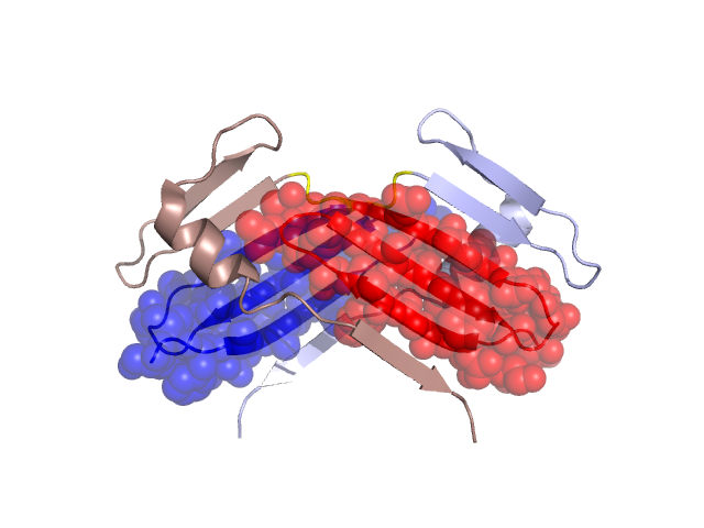
Structural Details of PDB entry 1MBY
Structural Details of PDB entry 1MBY
PDBid Chains Hinge Swapped Domain
1MBY
A,B
A:877-879,B:877-879
A:845-876,B:845-876
Swapped-domain interface residues and interactions:
Chains Residues
A
846 , 848 , 853 , 864 , 866 , 868 , 869 , 870 , 871 , 872 , 873 , 874 , 875 , 876 , 877 , 878 , 879 , 880 , 881 , 882 , 883 , 884 , 886 ,
B
846 , 848 , 853 , 864 , 866 , 868 , 869 , 870 , 871 , 872 , 873 , 874 , 875 , 876 , 877 , 878 , 879 , 880 , 881 , 882 , 883 , 884 , 886 ,
Non-swapped-domain interface residues and interactions:
Mutations in critical regions:
Chains
Hinge
Domain swapped interface Non-swapped interface Swapped Domain
A No mutation MET(874)VAL, LEU(909)CYS, MET(874)VAL, B No mutation MET(874)VAL, LEU(909)CYS, MET(874)VAL,
HIDE output:
JMOL Visualization:
2D-plot:
JOY Structural annotation for hinge hinge and swapped domain:
JOY output: