Pfam Domains mapped on to the structure: 1L6S
No.
Chain ID
Pfam ID
Pfam Description
Linkout - Pfam
Linkout - CDD
1
A
PF00490
Delta-aminolevulinic acid dehydratase
PF00490
PF00490
Conserved Domain Database Superfamily Annotations: 1L6S
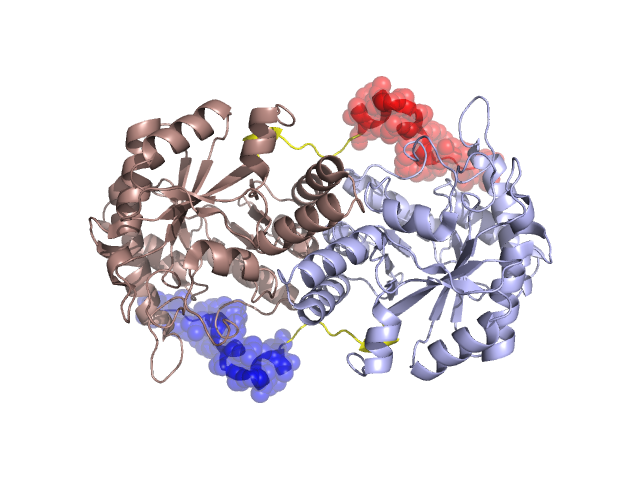
Structural Details of PDB entry 1L6S
Structural Details of PDB entry 1L6S
PDBid Chains Hinge Swapped Domain
1L6S
A,B
A:21-29,B:21-29
A:1-20,B:1-20
Swapped-domain interface residues and interactions:
Chains Residues
A
1 , 3 , 5 , 6 , 7 , 8 , 11 , 12 , 17 , 20 , 140 , 168 , 169 , 216 , 217 , 218 ,
B
1 , 3 , 5 , 6 , 7 , 8 , 11 , 12 , 17 , 20 , 140 , 168 , 169 , 216 , 217 , 218 ,
Non-swapped-domain interface residues and interactions:
Chains Residues
A
21 , 22 , 23 , 48 , 137 , 139 , 143 , 172 , 198 , 199 , 201 , 202 , 220 , 221 , 222 , 223 , 224 , 225 , 226 , 227 , 230 , 231 , 234 , 249 , 250 , 251 , 252 , 253 , 256 , 257 , 273 , 276 , 277 , 278 , 280 , 281 , 284 , 286 , 287 , 291 , 292 , 295 , 296 , 298 , 299 , 302 , 303 , 323 ,
B
21 , 22 , 23 , 48 , 137 , 139 , 143 , 172 , 198 , 199 , 201 , 202 , 220 , 221 , 222 , 223 , 224 , 225 , 226 , 227 , 230 , 231 , 234 , 249 , 250 , 251 , 252 , 253 , 256 , 257 , 273 , 276 , 277 , 278 , 280 , 281 , 284 , 286 , 287 , 291 , 292 , 295 , 296 , 298 , 299 , 302 , 303 ,
Mutations in critical regions:
Chains
Hinge
Domain swapped interface Non-swapped interface Swapped Domain
A No mutation No mutation No mutation No mutation B No mutation No mutation No mutation No mutation
HIDE output:
JMOL Visualization:
2D-plot:
JOY Structural annotation for hinge hinge and swapped domain:
JOY output: