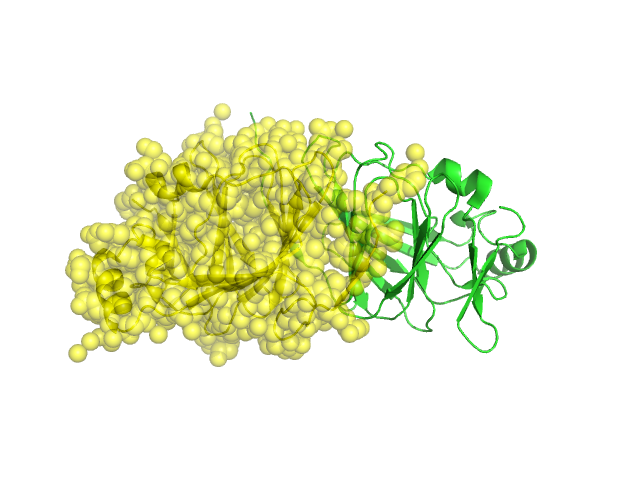
Structural Details of PDB entry 1J3Q
Structural Details of PDB entry 1J3Q
PDBid Chains Hinge Swapped Domain
1J3Q
A,B
A:-,B:-
A:1--1,B:3--1
Swapped-domain interface residues and interactions:
Non-swapped-domain interface residues and interactions:
Chains Residues
A
1 , 3 , 4 , 5 , 6 , 7 , 8 , 9 , 10 , 11 , 13 , 60 , 64 , 65 , 66 , 67 , 69 , 95 , 96 , 97 , 99 , 101 , 103 , 113 , 114 , 115 , 119 , 121 , 124 , 125 , 126 , 127 , 128 , 130 , 132 , 133 , 134 , 135 , 150 , 152 , 154 , 187 ,
B
3 , 4 , 5 , 6 , 7 , 8 , 9 , 10 , 11 , 13 , 60 , 63 , 64 , 65 , 66 , 67 , 69 , 95 , 96 , 97 , 99 , 101 , 103 , 113 , 114 , 115 , 117 , 119 , 121 , 124 , 125 , 126 , 127 , 128 , 130 , 132 , 133 , 134 , 135 , 150 , 152 , 154 ,