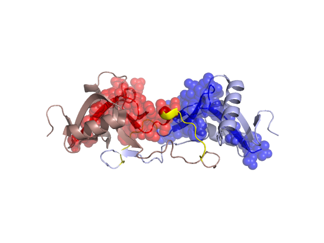
Structural Details of PDB entry 1J34
Structural Details of PDB entry 1J34
PDBid Chains Hinge Swapped Domain
1J34
A,B
A:89-96,B:285-292
A:97-129,B:293-323
Swapped-domain interface residues and interactions:
Chains Residues
A
93 , 94 , 95 , 98 , 100 , 101 , 103 , 113 , 114 , 115 , 116 , 129 ,
B
289 , 290 , 291 , 295 , 297 , 304 , 305 , 306 , 307 , 308 ,
Non-swapped-domain interface residues and interactions:
Chains Residues
A
27 , 38 , 41 , 42 , 43 , 44 , 45 , 48 , 69 , 70 , 71 , 72 , 73 , 78 , 79 , 80 , 83 , 84 , 85 , 87 , 89 , 91 ,
B
227 , 238 , 241 , 242 , 243 , 244 , 245 , 248 , 267 , 268 , 269 , 271 , 272 , 274 , 275 , 276 , 277 , 278 , 279 , 280 , 281 , 283 , 285 , 287 ,