Pfam Domains mapped on to the structure: 1I6O
No.
Chain ID
Pfam ID
Pfam Description
Linkout - Pfam
Linkout - CDD
1
A
PF00484
Carbonic anhydrase
PF00484
PF00484
Conserved Domain Database Superfamily Annotations: 1I6O
Structural Details of PDB entry 1I6O
Structural Details of PDB entry 1I6O
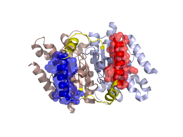
PDBid Chains Hinge Swapped Domain
1I6O
A,B
A:21-37,B:21-37
A:3-20,B:2-20
Swapped-domain interface residues and interactions:
Chains Residues
A
3 , 4 , 7 , 8 , 10 , 11 , 12 , 14 , 15 , 19 , 52 , 53 , 54 , 185 , 186 , 187 , 188 ,
B
2 , 3 , 4 , 7 , 8 , 10 , 11 , 12 , 14 , 15 , 19 , 52 , 53 , 54 , 185 , 186 , 187 , 188 ,
Non-swapped-domain interface residues and interactions:
Chains Residues
A
25 , 26 , 29 , 30 , 32 , 33 , 35 , 37 , 43 , 44 , 45 , 46 , 47 , 48 , 50 , 51 , 55 , 57 , 58 , 59 , 60 , 61 , 62 , 63 , 64 , 65 , 66 , 75 , 76 , 79 , 80 , 82 , 83 , 87 , 92 , 102 , 103 , 113 , 115 , 116 , 118 , 119 , 163 , 177 , 179 , 183 , 184 , 190 , 215 ,
B
25 , 26 , 29 , 30 , 35 , 37 , 43 , 44 , 45 , 46 , 47 , 48 , 50 , 51 , 55 , 57 , 58 , 60 , 61 , 62 , 63 , 64 , 65 , 66 , 74 , 75 , 76 , 79 , 80 , 82 , 83 , 87 , 92 , 94 , 102 , 103 , 113 , 115 , 116 , 118 , 119 , 163 , 177 , 179 , 183 , 184 , 190 ,
Mutations in critical regions:
Chains
Hinge
Domain swapped interface Non-swapped interface Swapped Domain
A No mutation No mutation No mutation No mutation B No mutation No mutation No mutation No mutation
HIDE output:
JMOL Visualization:
2D-plot:
JOY Structural annotation for hinge hinge and swapped domain:
JOY output: