Home BrowseSearch Help Group

Pfam Domains mapped on to the structure: 1I5E

No. Chain ID Pfam ID Pfam Description Linkout - Pfam Linkout - CDD
1 A PF00156 Phosphoribosyl transferase domain PF00156 PF00156

Conserved Domain Database Superfamily Annotations: 1I5E

No. PDB ID PSSM ID CDD Accession Superfamily Short Name Linkout - CDD
1 1I5E 206754 cd06223 PRTases_typeI - cl00309
2 1I5E 212186 cl00309 PRTases_typeI superfamily - -
Structural Details of PDB entry 1I5E

Structural Details of PDB entry 1I5E

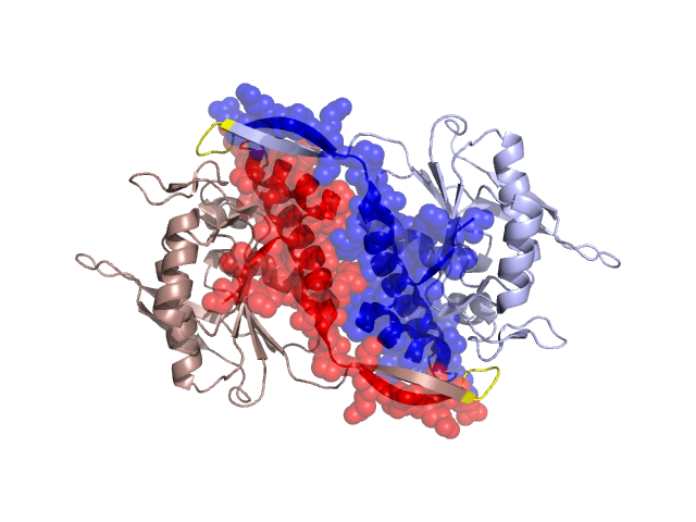
PDBid Chains Hinge Swapped Domain
1I5E A,B A:57-61,B:57-61 A:2-56,B:2-56

Swapped-domain interface residues and interactions:

Chains Residues
A 9, 10, 11, 13, 14, 17, 20, 21, 22, 39, 42, 43, 45, 46, 48, 50, 55, 56, 57, 58, 64, 65, 66, 67,
B 9, 10, 11, 13, 14, 17, 20, 21, 22, 39, 42, 43, 45, 46, 48, 50, 55, 56, 57, 58, 64, 65, 66, 67,

Non-swapped-domain interface residues and interactions:

Chains Residues
A 59, 60, 62, 91, 189, 193, 195, 196, 199, 209,
B 59, 60, 62, 91, 189, 195, 196, 199,

Swapped domains are represented using trasperent spheres.
Non-swapped part is represented using light color and cartoon representation.
Hinge region is shown in yellow color.

Mutations in critical regions:

Chains Hinge Domain swapped interface Non-swapped interface Swapped Domain
ANo mutationNo mutationNo mutationNo mutation
BNo mutationNo mutationNo mutationNo mutation

HIDE output:

Homologues found through HIDE algorithm

JMOL Visualization:

Jmol_logo            Jmol_logo            Jmol_logo

2D-plot:

A:1I5E B:1I5E

JOY Structural annotation for hinge hinge and swapped domain:

Hinge Swapped domain

JOY output:

ali file:1I5E.ali
atm file:1I5E.atm
cof file:1I5E.cof
hbd file:1I5E.hbd
html file:1I5E.html
pdb file:1I5E.pdb
ps file:1I5E.ps
psa file:1I5E.psa
rtf file:1I5E.rtf
sst file:1I5E.sst
tem file:1I5E.tem