Home BrowseSearch Help Group

Pfam Domains mapped on to the structure: 1HT9

No. Chain ID Pfam ID Pfam Description Linkout - Pfam Linkout - CDD
1 A PF01023 S-100/ICaBP type calcium binding domain PF01023 PF01023
2 A PF13499 EF-hand domain pair PF13499 PF13499
3 A PF00036 EF hand PF00036 PF00036

Conserved Domain Database Superfamily Annotations: 1HT9

No. PDB ID PSSM ID CDD Accession Superfamily Short Name Linkout - CDD
1 1HT9 88292 cd00213 S-100 - cl08302
2 1HT9 208857 cl08302 EFh superfamily - -
Structural Details of PDB entry 1HT9

Structural Details of PDB entry 1HT9

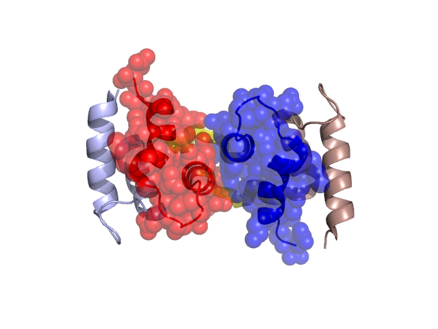
PDBid Chains Hinge Swapped Domain
1HT9 A,B A:38-45,B:38-45 A:46-75,B:46-75

Swapped-domain interface residues and interactions:

Chains Residues
A 22, 23, 24, 25, 45, 46, 47, 48, 49, 50, 51, 52, 53, 59, 60, 61, 62, 63, 66, 69, 70, 72, 73, 74, 75,
B 1, 3, 6, 22, 23, 24, 25, 46, 47, 48, 49, 50, 51, 52, 53, 59, 60, 61, 62, 63, 66, 69, 70, 72, 73,

Non-swapped-domain interface residues and interactions:

Chains Residues
A 0, 1, 6, 7, 10, 19, 28, 29, 31, 32, 36, 39, 40, 43,
B 7, 10, 19, 28, 29, 31, 32, 36, 39, 40, 43, 45,

Swapped domains are represented using trasperent spheres.
Non-swapped part is represented using light color and cartoon representation.
Hinge region is shown in yellow color.

Mutations in critical regions:

Chains Hinge Domain swapped interface Non-swapped interface Swapped Domain
AMET(43)PRO, ASN(22)GLN, MET(43)PRO, No mutation
BMET(43)PRO, ASN(22)GLN, MET(43)PRO, No mutation

HIDE output:

Homologues found through HIDE algorithm

JMOL Visualization:

Jmol_logo            Jmol_logo            Jmol_logo

2D-plot:

A:1HT9 B:1HT9

JOY Structural annotation for hinge hinge and swapped domain:

Hinge Swapped domain

JOY output:

ali file:1HT9.ali
atm file:1HT9.atm
cof file:1HT9.cof
hbd file:1HT9.hbd
html file:1HT9.html
pdb file:1HT9.pdb
ps file:1HT9.ps
psa file:1HT9.psa
rtf file:1HT9.rtf
sst file:1HT9.sst
tem file:1HT9.tem