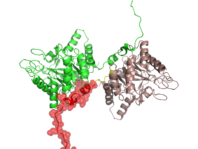
Structural Details of PDB entry 1H7N
Structural Details of PDB entry 1H7N
PDBid Chains Hinge Swapped Domain
1H7N
A,B
A:33-40,B:33-40
A:1-32,B:1-32
Swapped-domain interface residues and interactions:
Chains Residues
A
11 , 12 , 14 , 17 , 19 , 20 , 21 , 23 , 24 , 26 , 27 , 29 , 30 , 31 , 32 ,
D
199 , 200 , 201 , 203 , 256 , 258 , 283 ,
Non-swapped-domain interface residues and interactions:
Chains Residues
A
33 , 34 , 35 , 37 , 39 , 40 , 84 , 85 , 125 , 127 , 174 , 187 , 190 , 191 , 194 , 199 , 200 , 201 , 203 , 254 , 256 , 258 , 280 , 283 , 324 , 325 , 340 ,
D
11 , 12 , 14 , 17 , 19 , 20 , 21 , 23 , 24 , 26 , 27 , 29 , 30 , 31 , 32 , 33 , 34 , 35 , 37 , 39 , 40 , 84 , 85 , 125 , 127 , 174 , 187 , 190 , 191 , 194 , 254 , 280 , 324 , 325 ,