Pfam Domains mapped on to the structure: 1H4V
No.
Chain ID
Pfam ID
Pfam Description
Linkout - Pfam
Linkout - CDD
1
B
PF03129
Anticodon binding domain
PF03129
PF03129
Conserved Domain Database Superfamily Annotations: 1H4V
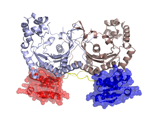
Structural Details of PDB entry 1H4V
Structural Details of PDB entry 1H4V
PDBid Chains Hinge Swapped Domain
1H4V
A,B
A:321-330,B:321-330
A:331-421,B:331-421
Swapped-domain interface residues and interactions:
Chains Residues
A
29 , 30 , 33 , 34 , 35 , 247 , 341 , 342 , 343 , 345 , 346 , 349 , 352 , 353 , 356 , 358 , 359 , 361 , 376 , 421 ,
B
29 , 30 , 33 , 34 , 35 , 247 , 341 , 342 , 343 , 345 , 346 , 349 , 352 , 353 , 356 , 358 , 359 , 361 , 376 ,
Non-swapped-domain interface residues and interactions:
Chains Residues
A
2 , 3 , 4 , 5 , 6 , 7 , 9 , 10 , 11 , 12 , 13 , 14 , 17 , 21 , 28 , 32 , 37 , 38 , 39 , 40 , 42 , 43 , 44 , 45 , 46 , 48 , 49 , 63 , 66 , 68 , 70 , 71 , 74 , 76 , 78 , 88 , 91 , 92 , 93 , 98 , 99 , 101 , 103 , 109 , 111 , 113 , 115 , 116 , 122 , 123 , 136 , 138 , 142 , 145 , 146 , 149 , 153 , 242 , 243 , 246 , 295 , 296 , 326 ,
B
2 , 3 , 4 , 5 , 6 , 7 , 9 , 10 , 11 , 12 , 13 , 14 , 17 , 21 , 28 , 32 , 37 , 38 , 39 , 40 , 42 , 43 , 44 , 45 , 46 , 48 , 49 , 63 , 66 , 68 , 70 , 71 , 74 , 76 , 78 , 88 , 91 , 92 , 93 , 98 , 99 , 101 , 103 , 109 , 111 , 113 , 115 , 116 , 122 , 123 , 136 , 138 , 142 , 145 , 146 , 149 , 153 , 242 , 243 , 246 , 295 , 296 , 326 ,
Mutations in critical regions:
Chains
Hinge
Domain swapped interface Non-swapped interface Swapped Domain
A No mutation No mutation No mutation No mutation B No mutation No mutation No mutation No mutation
HIDE output:
JMOL Visualization:
2D-plot:
JOY Structural annotation for hinge hinge and swapped domain:
JOY output: