Home BrowseSearch Help Group

Pfam Domains mapped on to the structure: 1H2D

No. Chain ID Pfam ID Pfam Description Linkout - Pfam Linkout - CDD
1 A PF07447 Matrix protein VP40 PF07447 PF07447

Conserved Domain Database Superfamily Annotations: 1H2D

No. PDB ID PSSM ID CDD Accession Superfamily Short Name Linkout - CDD
1 1H2D 116068 VP40 superfamily C -
Structural Details of PDB entry 1H2D

Structural Details of PDB entry 1H2D

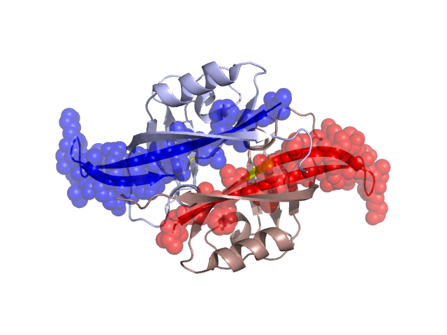
PDBid Chains Hinge Swapped Domain
1H2D A,B A:95-96,B:95-96 A:69-94,B:69-94

Swapped-domain interface residues and interactions:

Chains Residues
A 70, 72, 74, 76, 78, 91, 93, 164, 187, 189, 190,
B 69, 70, 72, 74, 76, 90, 91, 92, 93, 187, 188, 189, 190,

Non-swapped-domain interface residues and interactions:

Chains Residues
A 95, 97, 99, 100, 102, 104, 130, 146, 148, 151, 155, 158, 160, 161, 165, 178, 180, 182, 184, 186,
B 95, 97, 99, 100, 102, 104, 146, 148, 151, 155, 158, 160, 161, 164, 165, 182, 184, 186, 191,

Swapped domains are represented using trasperent spheres.
Non-swapped part is represented using light color and cartoon representation.
Hinge region is shown in yellow color.

Mutations in critical regions:

Chains Hinge Domain swapped interface Non-swapped interface Swapped Domain
ANo mutationNo mutationNo mutationNo mutation
BNo mutationNo mutationNo mutationNo mutation

HIDE output:

Homologues found through HIDE algorithm

JMOL Visualization:

Jmol_logo            Jmol_logo            Jmol_logo

2D-plot:

A:1H2D B:1H2D

JOY Structural annotation for hinge hinge and swapped domain:

Hinge Swapped domain

JOY output:

ali file:1H2D.ali
atm file:1H2D.atm
cof file:1H2D.cof
hbd file:1H2D.hbd
html file:1H2D.html
pdb file:1H2D.pdb
ps file:1H2D.ps
psa file:1H2D.psa
rtf file:1H2D.rtf
sst file:1H2D.sst
tem file:1H2D.tem