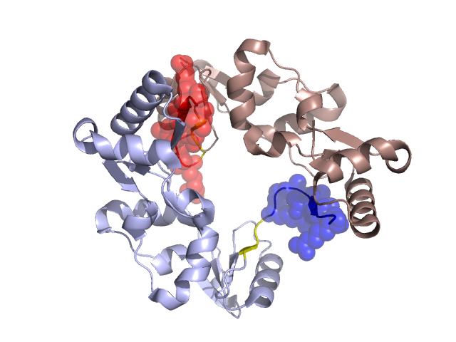
Structural Details of PDB entry 1GXJ
Structural Details of PDB entry 1GXJ
PDBid Chains Hinge Swapped Domain
1GXJ
A,B
A:646-649,B:646-649
A:650-658,B:650-658
Swapped-domain interface residues and interactions:
Chains Residues
A
557 , 560 , 561 , 566 , 567 , 568 , 569 , 570 , 571 , 650 , 651 , 652 , 653 , 654 , 655 , 656 , 657 , 658 ,
B
557 , 560 , 561 , 564 , 566 , 567 , 568 , 569 , 570 , 575 , 576 , 577 , 651 , 652 , 653 , 654 , 655 , 656 ,
Non-swapped-domain interface residues and interactions:
Chains Residues
A
553 , 564 , 572 , 623 , 624 , 627 , 630 , 631 , 643 , 644 , 645 ,
B
501 , 550 , 553 , 554 , 572 , 574 , 579 , 612 , 615 ,