Pfam Domains mapped on to the structure: 1GAF
Conserved Domain Database Superfamily Annotations: 1GAF
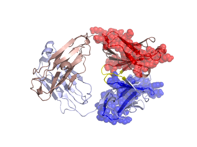
Structural Details of PDB entry 1GAF
Structural Details of PDB entry 1GAF
PDBid Chains Hinge Swapped Domain
1GAF
H,L
H:41-45,L:40-44
H:1-40,L:1-39
Swapped-domain interface residues and interactions:
Non-swapped-domain interface residues and interactions:
Chains Residues
H
44 , 45 , 47 , 50 , 59 , 61 , 62 , 95 , 100 , 101 , 102 , 104 , 123 , 124 , 125 , 126 , 129 , 130 , 136 , 138 , 142 , 144 , 165 , 166 , 167 , 168 , 170 , 180 , 182 , 184 , 215 ,
L
42 , 44 , 46 , 49 , 55 , 87 , 89 , 91 , 94 , 95 , 96 , 98 , 100 , 116 , 118 , 119 , 121 , 123 , 124 , 131 , 133 , 135 , 137 , 138 , 160 , 162 , 163 , 164 , 167 , 174 , 176 , 180 , 209 , 212 , 214 ,
Mutations in critical regions:
Chains
Hinge
Domain swapped interface Non-swapped interface Swapped Domain
H No mutation No mutation No mutation No mutation L ASP(41)GLY, GLY(42)LYS, ILE(44)PRO, LEU(36)TYR, GLY(42)LYS, ILE(44)PRO, ARG(46)LEU, HIS(55)GLU, LEU(89)GLN, TYR(94)LEU, ARG(96)LEU, LEU(15)VAL, GLU(17)ASP, SER(20)THR, LEU(21)ILE, ARG(24)GLN, GLU(28)ASP, GLY(31)ASN, GLY(34)ASN, LEU(36)TYR,
HIDE output:
JMOL Visualization:
2D-plot:
JOY Structural annotation for hinge hinge and swapped domain:
JOY output: